Atrial oversensing by the ventricular lead
Tracing
Manufacturer Medtronic
Device ICD
Field Oversensing
N° 23
Patient
Patient with ischemic cardiomyopathy implanted with a dual-chamber ICD (Evera XT DR), hospitalized for lipothymias; history of paroxysmal AF; this tracing illustrates the problem of atrial oversensing by the ventricular lead.
Graph and trace
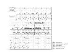
On the graph, there is a regular and fast atrial activity and a railroad track pattern at the level of the ventricular intervals with a very short interval (at the limit of the programmed blanking value) and a longer interval; at the end of the tracing, over a few intervals, very fast ventricular rate superimposed on the atrial activity.
- the EGM confirms the presence of a regular atrial tachycardia with intervals at 220-230 ms;
- correct sensing of the QRS complex;
- oversensing at the ventricular channel of a signal corresponding to atrial activity; due to the adaptive ventricular sensitivity, the atrial signals of equivalent amplitude are not sensed by the ventricular lead, the device being less sensitive at the beginning of the interval and the sensitivity adapting to the amplitude of the QRS complex;
- sensing of the ensuing QRS complex classified as FS;
- alternation between QRS complex detection and oversensing at the end of diastole (maximum sensitivity);
- on this interval, absence of spontaneous QRS complex, persistence of oversensing (intervals classified as FS), the device remaining extremely sensitive (level of sensitivity remaining adapted to the oversensed low-amplitude atrial signal); ventricular pause possibly explaining the lipothymias;
- end of oversensing following the occurrence of a QRS complex.
Other articles that may be of interest to you
EGM recordings
EGM recordings

This tracing thus shows an example of atrial activity oversensing by the ventricular canal during an episode of atrial tachycardia. The graph is characteristic of an oversensing of a physiological signal (P-wave, T-wave, R-wave double counting) with a railroad track pattern and an alternation between long and short ventricular intervals. The ventricular EGM shows an alternation between 2 signals of different morphology, the first corresponding obviously to the QRS complex, the second to an oversensing of atrial activity. Oversensing issues are of particular concern in dependent patients since
1) they may cause ventricular pauses,
2) the oversensing may be prolonged in the absence of spontaneous QRS complexes, the level of sensitivity remaining maximal and adapted to the amplitude of the oversensed signals which is often low.
Oversensing of atrial depolarization by the right ventricular lead is rare and occurs mainly in patients implanted with an integrated bipolar lead. In patients in sinus rhythm, the right ventricular lead senses both atrial and ventricular depolarizations, with the PR interval being longer than the ventricular blanking period. If the patient has a complete atrioventricular block, P-wave oversensing may inhibit ventricular pacing and lead to asystole. Similarly, oversensing of atrial depolarization during atrial flutter or atrial tachycardia can cause both inappropriate therapies and asystole if the patient is dependent.
Atrial oversensing by the ventricular lead preferentially occurs in 2 clinical situations:
Atrial oversensing may also be observed under more exceptional circumstances:
Several solutions can be considered to address the problem and avoid the occurrence of inappropriate therapies or ventricular pauses in a dependent patient: