Specific algorithm for diagnosing T-wave oversensing
Tracing
Manufacturer Medtronic
Device ICD
Field Oversensing
N° 21
Patient
Young patient implanted with a single-chamber ICD (Protecta XT VR) for Brugada syndrome; this tracing illustrates the value of a specific algorithm for diagnosing T-wave oversensing and avoiding the occurrence of inappropriate therapies.
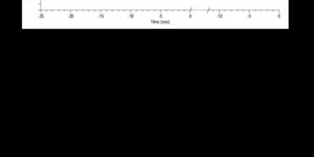
Graph and trace
On this graph, one can observe that the device rendered the diagnosis of T-wave oversensing which suspended the therapy.
- the EGM demonstrates an oversensing of the T-wave after an R-wave of very low amplitude;
- following a ventricular extrasystole, the ensuing low-amplitude QRS complex is undersensed;
- when the oversensing of the T-wave is maintained over several intervals, the VF counter is filled although no therapy is delivered, the device latter having diagnosed the oversensing;
- analysis by the algorithm continues on sliding windows with, from cycle to cycle, a diagnosis of T-wave oversensing.
Other articles that may be of interest to you
EGM recordings
EGM recordings

The crucial step in minimizing T-wave oversensing is the choice of bandpass filters which vary according to manufacturers (difference in terms of high-pass and low-pass filters). The slew rate, characteristic of a T-wave (smooth signal), is typically less than that of an R-wave (sharper signal) with a usual frequency of less than 5 Hz, although may be altered under various conditions (drugs, ischemia, sympathetic tone, metabolic abnormalities). In certain situations, the filters and sensing amplifiers may not provide a sufficiently wide difference in terms of absolute value of the R-wave and T-wave amplitudes, such that the amplitude of the T-wave may exceed the detection threshold.
T-wave oversensing following a spontaneous ventricular beat preferentially occurs in the presence of a low-amplitude R wave as in the case of this patient. The sensitivity and gain being automatically adjusted to the amplitude of the immediate R-wave detected, when the R-wave is of low amplitude, the device quickly reaches high levels of sensitivity. Reprogramming possibilities are limited upon occurrence of this type of problem (no possibility of modifying the filters or of reprogramming the adaptation level or delay on the MedtronicTM devices, and no programming margin for ventricular sensitivity). These situations are therefore difficult, often lead to programming compromises and it may therefore be necessary to reposition the lead.
This tracing illustrates the interest of a specific algorithm whose aim is not to prevent T-wave oversensing but whose purpose is to detect its presence and avoid the occurrence of inappropriate therapies. The discrimination of R-waves and T-waves is achieved by means of a differential filter which amplifies the difference between the 2 signals. The algorithm recognizes the oversensing of the T-wave by identifying the repetition of sequences with alternation between two signals of variable frequencies (a sharp signal, a smooth signal) with fixed intervals (fixed RT intervals, fixed TR intervals). Various parameters are hence analyzed and if all are fulfilled on 6 consecutive intervals, the T-wave oversensing counter is incremented by +1. If a single criterion is not met, the set of 6 consecutive intervals is classified as normal. The next group of 6 events is then assessed using a rolling window. As long as 4 of the last 20 cycles fulfill the discrimination criterion of the T-wave, the device retains the diagnosis of T-wave oversensing and the therapy is not delivered. This algorithm is nominally programmed to ON.
This algorithm allows diagnosing T-wave oversensing and avoid the occurrence of inappropriate therapies in this setting. On the other hand, the problem which generated the oversensing, namely the low-amplitude R-wave, is not resolved. A change in the configuration of ventricular sensitivity arises and will be discussed in the following tracing. As a last resort, if no reprogramming option appears acceptable, repositioning of the lead may be proposed.