Single-chamber VVI mode
Tracing
Manufacturer Medtronic
Device PM
Field Pacing Modes
N° 5
Patient
Same patient as in tracing 1.
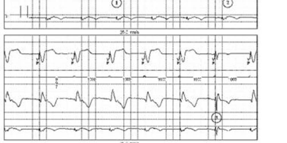
Graph and trace
Tracing 5a: programming in VVI mode 60 beats/minute;
- ventricular pacing at the base rate (1000 ms between VPs);
- 1:1 retrograde atrial conduction;
- fusion with spontaneous activation;
Tracing 5b: programming in VVI mode 40 beats/minute;
- spontaneous ventricular sensing and inhibition of ventricular pacing; functioning in sentinel mode.
Other articles that may be of interest to you




A single-chamber pacemaker operates in VVI mode when only one lead is positioned in the ventricle; the VVI mode can also be programmed in a dual-chamber pacemaker. The VVI mode provides single-chamber inhibited pacing at the programmed pacing rate, unless inhibited by a sensed event. Sensing applies only to the ventricle. An escape interval begins after any ventricular sensing or pacing. The first portion of the interval corresponds to the refractory period during which the pacemaker cannot sense a signal. A signal occurring in this refractory period does not recycle the escape interval. This refractory period is necessary to avoid double sensing of the same spontaneous or paced QRS complex. A signal sensed after this refractory period inhibits pacing and recycles the escape interval.
For this patient, programming of the base rate is essential. Indeed, at 60 beats per minute, the base rate is too high and a permanent ventricular pacing can be observed with inversion of the physiological atrioventricular activation sequence, retrograde conduction and pacemaker syndrome. Atrial contraction occurs almost synchronously with ventricular contraction while the atrioventricular valves are closed, causing a retrograde flow to the atria, pulmonary veins and vena cava. Pacemaker syndrome results from a complex combination of hemodynamic, neurohumoral and vascular alterations secondary to the loss of atrioventricular synchrony. A sometimes very disabling symptomatology due to increased atrial and venous pressures includes dyspnea, orthopnea, pulsations in the neck and in the chest, palpitations, or chest pain.
Conversely, at 40 beats/minute, the base rate is lower than the lowest spontaneous rate and the patient is not paced. This enables to: