Dual chamber discrimination: V=A followed by VT in the VF zone
Tracing
Manufacturer Abbott
Device ICD
Field Discrimination
N° 51
Patient
A 33-year old recipient of a Abbott dual chamber ICD implanted for multiples episodes of VT refractory to treatment with beta-adrenergic blockers, in the context of arrhythmogenic right ventricular dysplasia, was seen in consultation after receiving multiples shocks during exercise.
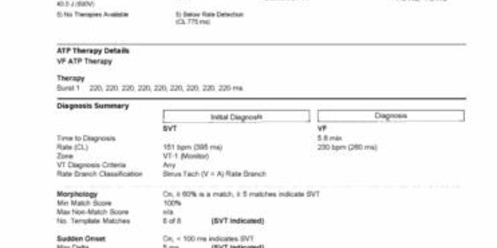
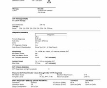
Main programmed parameters
- VF zone at 230 bpm, VT-2 zone at 176 bpm and VT-1 zone at 148 bpm VT
- 20 cycles in the VF zone, 20 cycles in the VT-2 zone and 20 cycles in the VT-1 zone were needed for the diagnosis
- Effective discrimination in the 2 VT zones
- V<A: if all criteria are fulfilled; morphology (60%, 5 out of 8), stability (80 ms), with 60-ms AV association delta, 12 intervals)
- V=A: if all criteria are fulfilled; morphology (60%, 5 out of 8), sudden onset (100 ms)
Graph and trace
Narrative
The episode was initially diagnosed as SVT in the V=A arm; in this patient, 1 out of 2 criteria were needed for the diagnosis of VT; both criteria indicate SVT, the device diagnosed SVT and, therefore, no therapy was delivered;
VF was subsequently diagnosed, 6 shocks were delivered before the cessation of therapy delivery without termination of the arrhythmia, during an episode that lasted over 5 minutes;
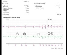
Tracing
- probable exercise-induced sinus tachycardia; the first cycle was classified as T1; the sudden onset criterion was analyzed from this cycle onward;
- from this complex onward, 8 cycles elapsed before the diagnosis, during which morphology was analyzed;
- diagnosis of SVT in the V=A arm (=SVT); the sudden and morphology criteria favor SVT;
- the episode continues with the acceleration of sinus tachycardia and remains diagnosed as SVT; redetection and analysis of the discrimination after each group of 6 cycles classified as T1;
- interruption of the EGM recording;
- ventricular extrasystole followed by acceleration of the tachycardia in the VF zone, with change in the morphology (0, x); retrograde conduction;
- diagnosis of VF after 20 cycles classified F; no discrimination in the VF zone;
- onset of the capacitors charge and delivery of burst during the charge;
- ineffective burst and continuation of the charge; confirmation of the persistence of the arrhythmia during the charge (underscored F);
- end of charge and delivery of electric shock;
- ineffective shock, redetection of VF (6 F cycles) and charge of the capacitors;
- second shock delivery;
- probable termination of the arrhythmia without diagnosis of return to sinus rhythm;
- early restart of the tachycardia; redetection of VF and charge of the capacitors;
- third shock delivery;
- redetection of VF;
- fourth shock delivery; probable termination, then early restart of the tachycardia;
- redetection of VF;
- fifth shock delivery;
- redetection of VF;
- delivery of 6th and last shock available for this episode;
- redetection of VF without remaining therapy available; the analysis of the intervals continues, though the cycles are no longer labeled F;
- end of 1:1 retrograde conduction; V>A, the diagnosis of VT is now ascertained;
- spontaneous termination of the arrhythmia and diagnosis of return to sinus rhythm.
Other articles that may be of interest to you


This tracing shows sinus tachycardia induced by vigorous exercise and initially accurately discriminated. The sudden onset and morphology parameters were both initially in favor of SVT. In this young patient presenting with arrhythmogenic right ventricular dysplasia, exercise prompted the onset of fast VT, detected in the VF zone. In this rate range, the defibrillator did not look for the origin of the arrhythmia and delivered a burst of ATP during capacitor charging. The several shocks delivered were either ineffective or followed by the immediate return of the tachyarrhythmia, sometimes observed due to the release of catecholamines in the context of this type of heart disease. After the delivery of 6 shocks, the delivery of therapies ended despite the persistence of the arrhythmia. The episode ended spontaneously after cessation of exercise. This case emphasizes the importance of appropriate programming that is patient/disease specific