Slow VT and unsuccessful burst
Tracing
Manufacturer Abbott
Device ICD
Field Therapy
N° 37
Patient
This 49-year-old man presenting with a very severe dilated cardiomyopathy and a 15% left ventricular ejection fraction, received an Atlas dual chamber ICD and was seen for a routine visit.
Main programmed parameters
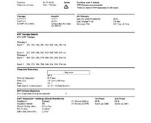
- VF zone at 207 bpm, VT-2 zone at 188 bpm, VT-1 zone at 102 bpm
- 12 cycles in the VF zone, 12 cycles in the VT-2 zone and 20 cycles in VT-1 zone were needed for the diagnosis
- Maximum sensitivity programmed at 0.3 mV
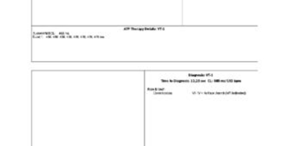
- VF zone: 1 shock at 25 J, followed by five 36-J shocks (maximum amplitude); VT-2 zone: 3 bursts followed by a single 20 J shock, followed by a single 32 J shock, followed by 2 shocks of maximal strength; VT-1 zone: 14 bursts of ATP followed by a single 5-J shock, followed by a single 10-J shock followed by two 15-J shocks
- Effective discrimination in the VT-1 zone
- DDI pacing mode at 50 bpm; DDI episode pacing mode; post-shock DDI pacing at 60 bpm
Graph and trace
Episode of VT at 102 bpm with 1 sequence of ATP; the heart rate after ATP is near the rate of the tachycardia, though is in the sinus zone.
Tracing
- This episode follows a previous, identical episode with erroneous diagnosis of restoration of sinus;
- Continuation of slow VT with AV dissociation;
- Diagnosis of VT-1 after 20 cycles T in the V>A arm;
- Burst of 9 stimuli;
- Continuation of the tachycardia; erroneous diagnosis of restoration of sinus rhythm due to slowing of the tachycardia.
Other articles that may be of interest to you


EGM recordings

The occurrence of slow VT (< 150 bpm) is relatively frequent in ICD recipients whose left ventricular ejection fraction is profoundly depressed. It may be useful to program initially a monitor zone for heart rates below 150 bpm, in order to detect the occurrence of this type of arrhythmia, which is often asymptomatic. If the episodes are short lived, the best strategy is probably to ignore them. If, however, the episodes are protracted and hemodynamically detrimental, it might be worthwhile to program a dedicated treatment zone. In that slow VT zone, various forms of ATP should be programmed. In a zone of slow VT, 1) one can increase the number of cycles needed to make the diagnosis in order to favor a spontaneous termination, 2) bursts should probably be preferred over ramps, as they are less aggressive; one must constantly keep in mind the risk of proarrhythmia by pacing, which can transform a slow, hemodynamically stable VT in a fast, life-threatening tachycardia, 3) one should not hesitate to program a large number of pacing sequences, 4) the programming of shocks is not mandatory and depends on the tolerability of the tachyarrhythmia.
In this patient, the burst was unsuccessful and this tracing illustrates the same issue as the previous tracing, namely a spurious diagnosis of restoration of sinus rhythm due to the slowing of the rate of the tachycardia.