
Chapter 4 · Oversensing · Case 4
Oversensing of the T wave on an older model
Patient and episode
Patient
- 41-year-old man with hypertrophic cardiomyopathy implanted with a single-chamber Atlas™ defibrillator for sustained ventricular tachycardia; routine consultatione.
Summary
- VT episode corresponding to the monitor zone
- VT episode corresponding to the monitor zone
The recording
Tap a number on the trace, or an entry in the list below
Episode 1
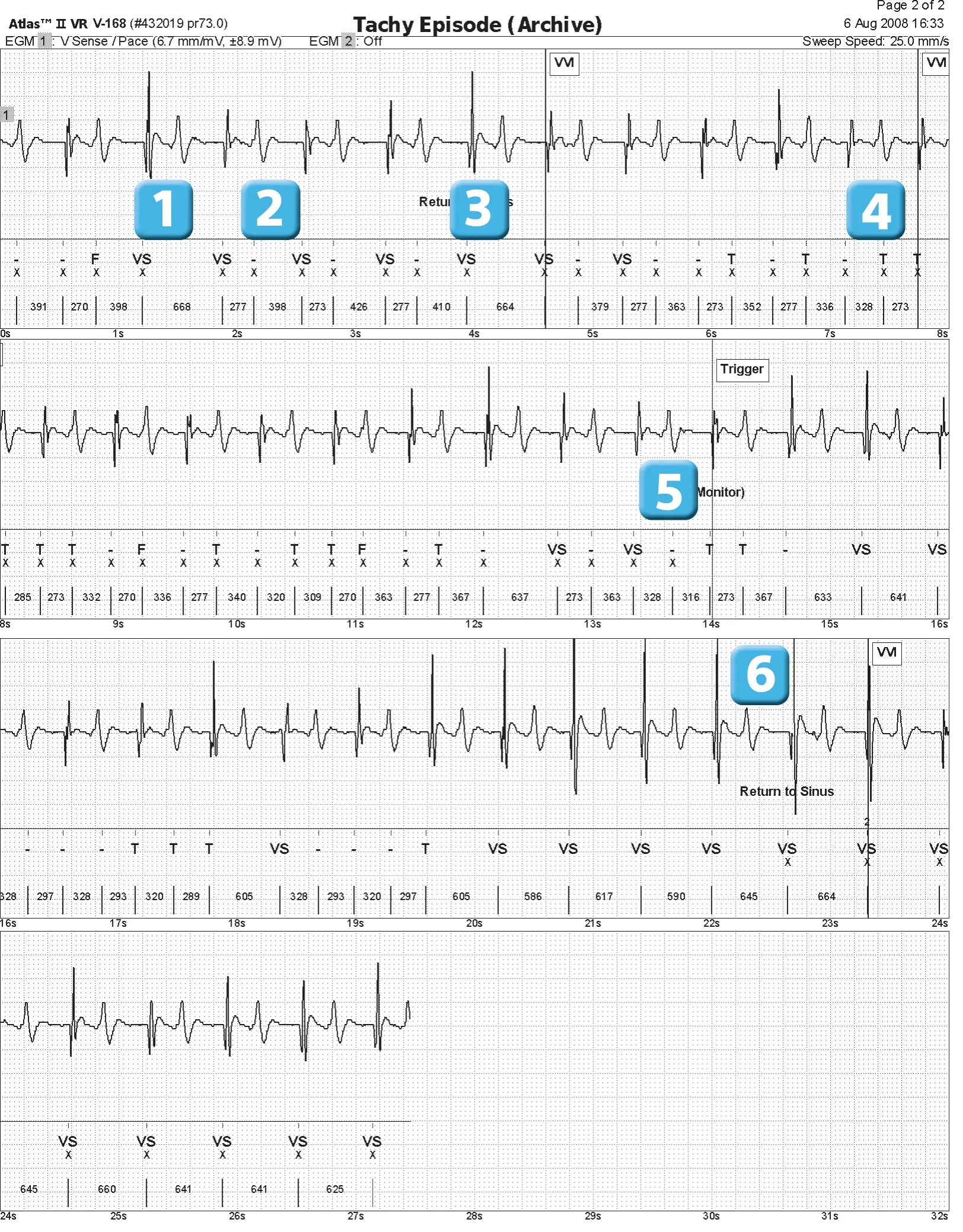
- Ventricle appearing to be of high amplitude without T wave oversensing; the channel present on the EGM corresponds to the V stim/detect channel: the signal displayed corresponds to the filtered and amplified signal on which the device performs its analysis; it is important to know the amplification (here 6.7 mm/mV) in order to estimate the amplitude of the different signals; remember that the measurement of an R wave corresponds to the measurement of the largest peak by comparing the positive and negative peaks of the signal
- ventricle with smaller amplitude and oversensing of the T wave (-); the amplitude of the T wave on this complex is more or less equivalent to that of the R wave
- diagnosis of sinus return after 5 consecutive VS or with unclassified intervals interspersed
- episode mode (VVI) after 4 classified T cycles following sinus return
- Repeated oversensing of the T wave and diagnosis of VT in the monitor zone (12 classified T cycles without diagnosis of sinus return); no therapy in this zone
- large variation in R wave amplitude; following ventricles of greater amplitude, no T wave oversensing; diagnosis of sinus retur
Episode 2
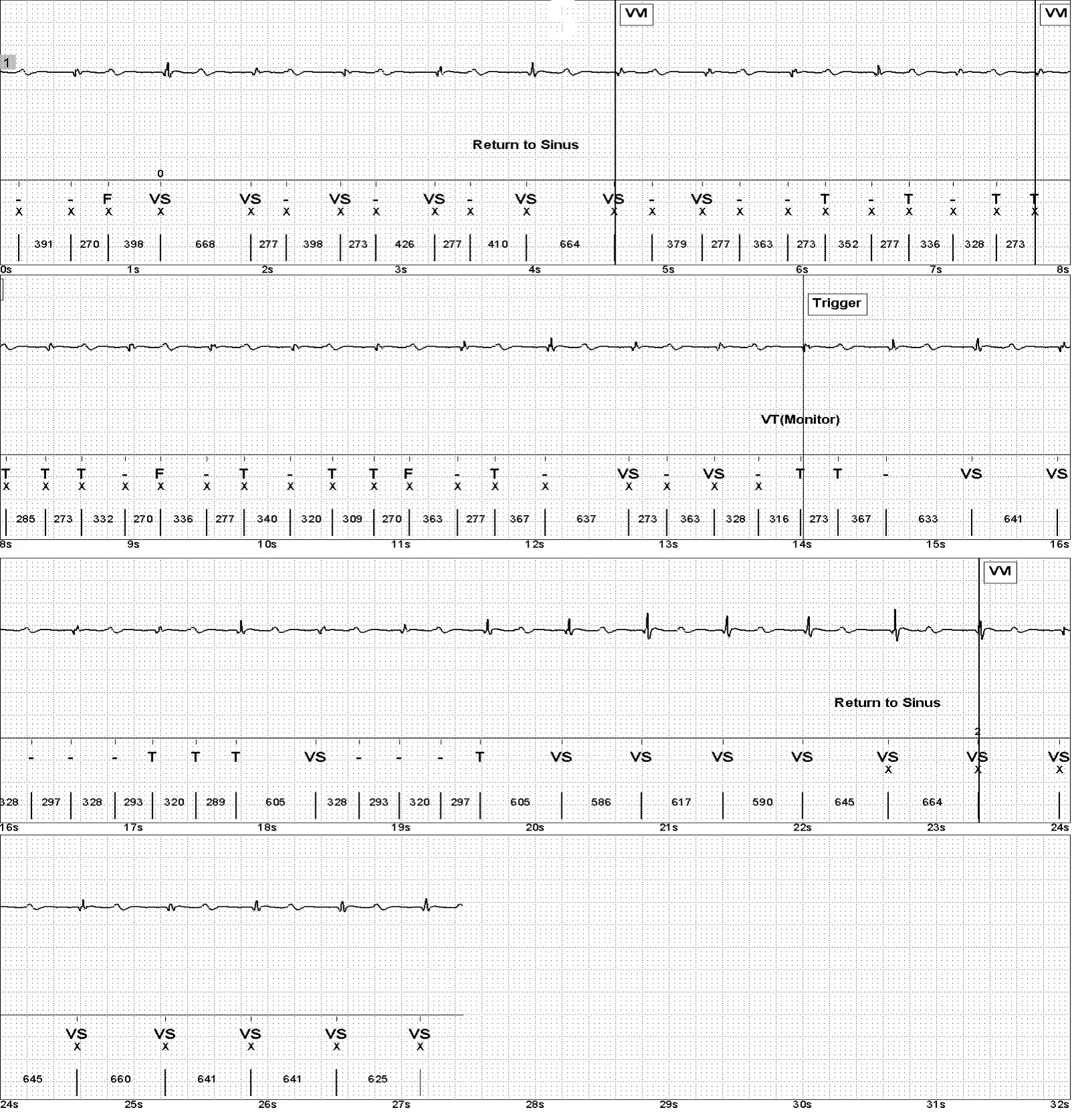
- This is the same episode and the same trace with a change in amplification; programming a gain of 1 mm/mV here provides a better understanding of the amplitude of the different signals and more clearly demonstrates the poor quality of ventricular sensing (very low amplitude signals).
Points to remember
- This trace was recorded on an Atlas™, an older model of defibrillator manufactured by Saint Jude Medical, and illustrates the problem of T wave oversensing, which at the time resulted in a significant number of inappropriate therapies.
- The systematic oversensing of a supernumerary cardiac signal results in the detection of two signals of different morphology (a high-frequency signal, the R wave, and a low-frequency signal, the T wave) for the same cardiac cycle, with alternation between two cycles (a short cycle and a longer cycle). For each cardiac cycle, the device counts both the R wave and the T wave as a second additional signal, resulting in a doubling of the heart rate.
- Oversensing of the T wave following a spontaneous ventricular contraction occurs preferentially in the presence of a low-amplitude R wave; sensitivity automatically adjusts to the amplitude of the preceding R wave: when the amplitude of the R wave is low, the device continuously operates at a high sensitivity level, which increases the risk of overdetecting the T wave.
- Oversensing of the T wave is promoted by exercise; exertion is sometimes associated with a decrease in the amplitude of the R wave and an increase in the amplitude of the T wave with an increase in signal velocity and therefore its slew rate, which causes the T waves to enter the detection bandwidth of ventricular events. Programming the number of cycles determining sinus return is very important for this type of problem because, in the presence of intermittent oversensing, this parameter has a very direct influence on the risk of inappropriate therapy.
- Abbott devices allow specific parameters to be programmed to cover and not detect the T wave: ventricular sensitivity, adaptation level, adaptation delay, and ventricular filters; however, the reprogramming options are limited if the main problem is the presence of a small-amplitude R wave
- in this patient, any reduction in ventricular sensitivity to avoid oversensing of the T wave would be associated with a major risk of undersensing of VF; in fact, the amplitude of the ventricular signal in sinus rhythm was measured at 2.6 mV; in addition, during exercise, as seen on this trace, the detection of ventriculograms is altered with a relatively large variation in amplitude from one ventricular complex to another
- a change in the adaptation level and adaptation delay made it possible to eliminate this oversensing of the T wave; a stress test confirmed the absence of any oversensing This patient’s situation remains a concern due to the poor quality of detection, and the question of repositioning the lead must be discussed.
Practice out loud
Just describe the tracing. What do you think is going on? Describing the numbers can help.
00:00
0%
—
From Implantable Cardioverter Defibrillator — clinical cases based on tracings by P. Bordachar, M. Strik, A. Thiyagarajah, S. Ploux. Published by Cardiocases. Every numbered marker on a recording is explained in the list beneath it; tap a marker on the trace or an entry in the list.