
Chapter 1 · Counters · Case 2
A ventricular arrhythmia in the VF zone
Patient and episode
Patient
- Patient with hypertrophic cardiomyopathy implanted with a single-chamber defibrillator (Visia AF VR) hospitalised for loss of consciousness.
The recording
Tap a number on the trace, or an entry in the list below
Rate / interval plot
EGM

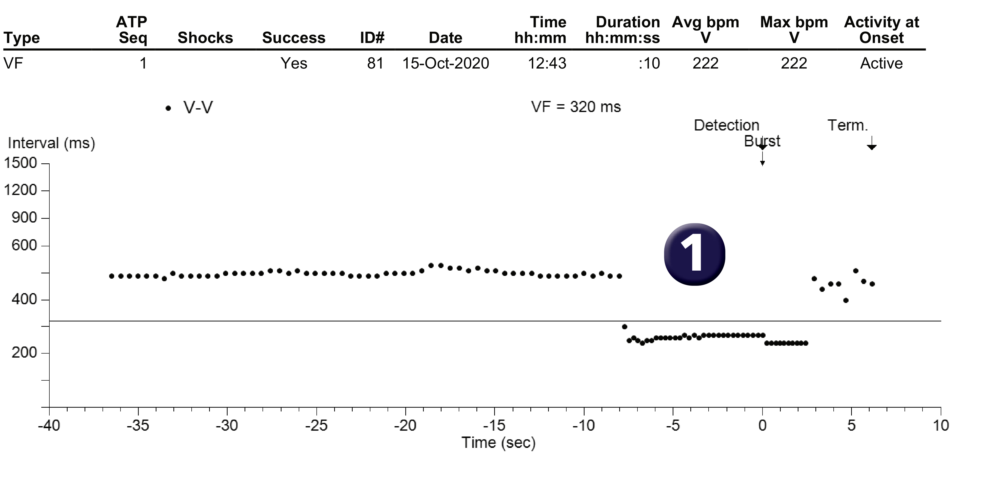
- What diagnosis is suggested by the interval plot? The episode was classified as VF by the device; a single detection zone was programmed (VF zone from 320 ms); the plot shows a sudden acceleration with regular ventricular cycles detected in the VF zone; a burst is delivered resulting in a slowing of the rate.
- What do EGM1 and EGM2 stand for? On this plot, we find EGM1 on the first line corresponding to bipolar ventricular sensing (RVtip to RVring), EGM2 on the second line corresponding to the shock channel (Can to Coil) and a third line with markers and intervals.
- What type of arrhythmia is it? This is a regular, monomorphic arrhythmia detected in the VF zone, probably corresponding to rapid VT.
- How is the initial detection counter in the VF zone programmed? The initial counter in the VF zone has been set to 30/40.
- What therapy is delivered? A burst before charging was delivered; the burst was effective and terminated the arrhythmia (the morphology of the QRS complexes was different from the morphology during tachycardia, reinforcing the presumed diagnosis of VT).
Points to remember
- In a double or triple chamber defibrillator, the comparison between atrial and ventricular activity sometimes provides certainty (ventricular rhythm faster than atrial rhythm) in terms of differentiating between ventricular arrhythmias and atrial arrhythmias; in a single-chamber defibrillator, certainty of the diagnosis is usually much more difficult; in this example, the diagnosis of VT is probable (different appearance of ventricular EGMs in tachycardia and post-termination, effectiveness of anti-tachycardia pacing, etc.) even if each criterion taken individually does not constitute definitive proof.
- By definition, ventricular fibrillation is a very rapid, irregular and polymorphic arrhythmia; it indicates the existence of chaotic ventricular activation, which becomes very disorganised; the diagnosis of VF made by a defibrillator is not an electrophysiological diagnosis (evidence of an irregular and polymorphic arrhythmia) but is based solely on an assessment of heart rate (faster than a programmable limit); in this example, this episode was classified as VF by the device even though it was clearly VT (regular and monomorphic arrhythmia) due to its rapidity.
- The observation that many patients record VT falling in the VF zone has led to a change in recommendations and programming of anti-tachycardia pacing as first-line treatment to reduce the number of shocks delivered.
Practice out loud
Just describe the tracing. What do you think is going on? Describing the numbers can help.
00:00
0%
—
From Medtronic ICD — clinical cases by P. Bordachar, A. Thiyagarajah, L. Fontagne, M. Strik, S. Ploux. Published by Cardiocases. Every numbered marker on a recording is explained in the list beneath it; tap a marker on the trace or an entry in the list.
Order hardcopyon Amazon