
Chapter 1 · Counters · Case 3
Utility of a probabilistic counter in the VF zone
Patient and episode
Patient
- Patient with hypertrophic cardiomyopathy implanted with a single-chamber defibrillator (Visia AF VR) hospitalised for loss of consciousness.
The recording
Tap a number on the trace, or an entry in the list below
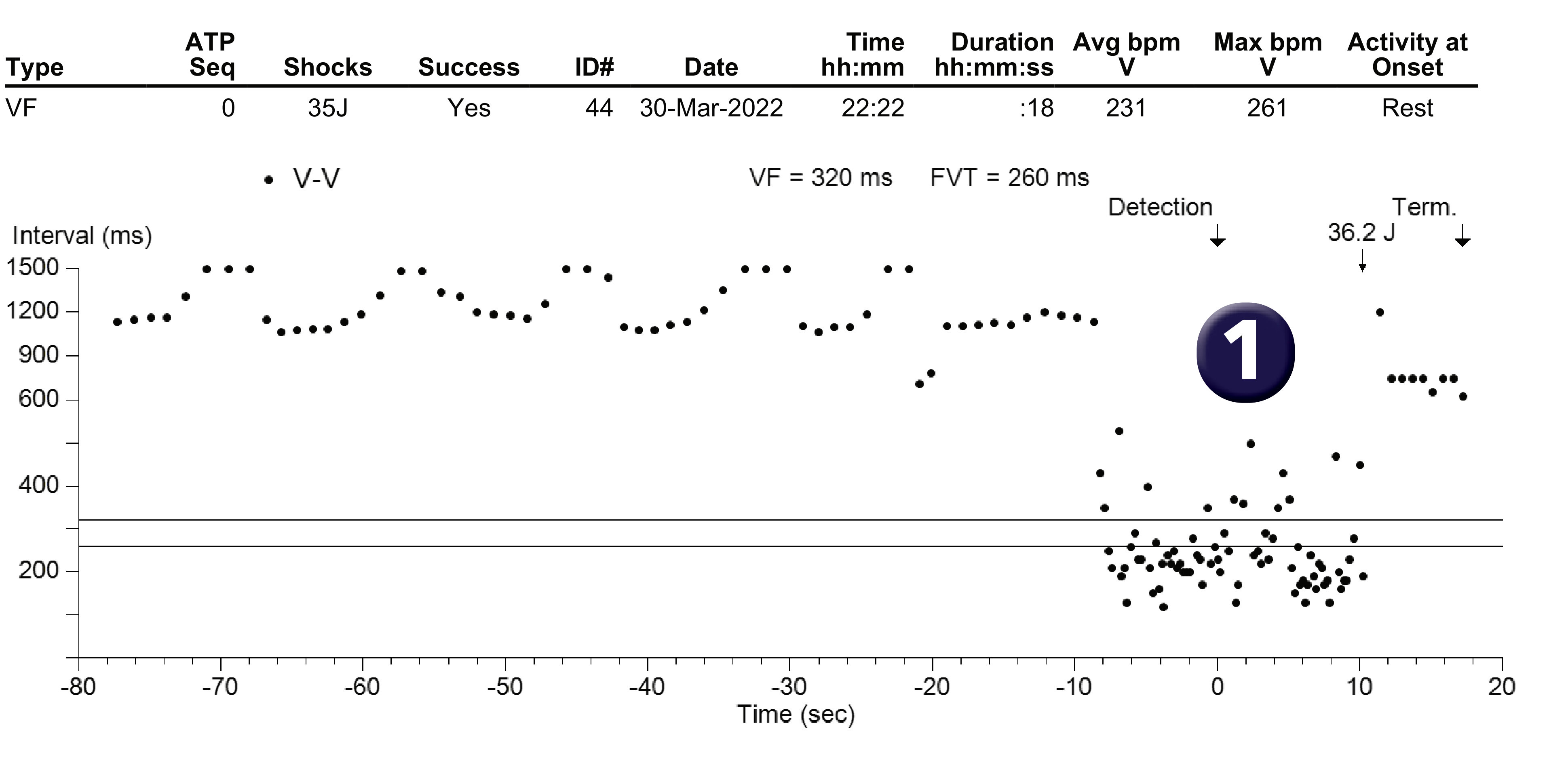
Rate / interval plot
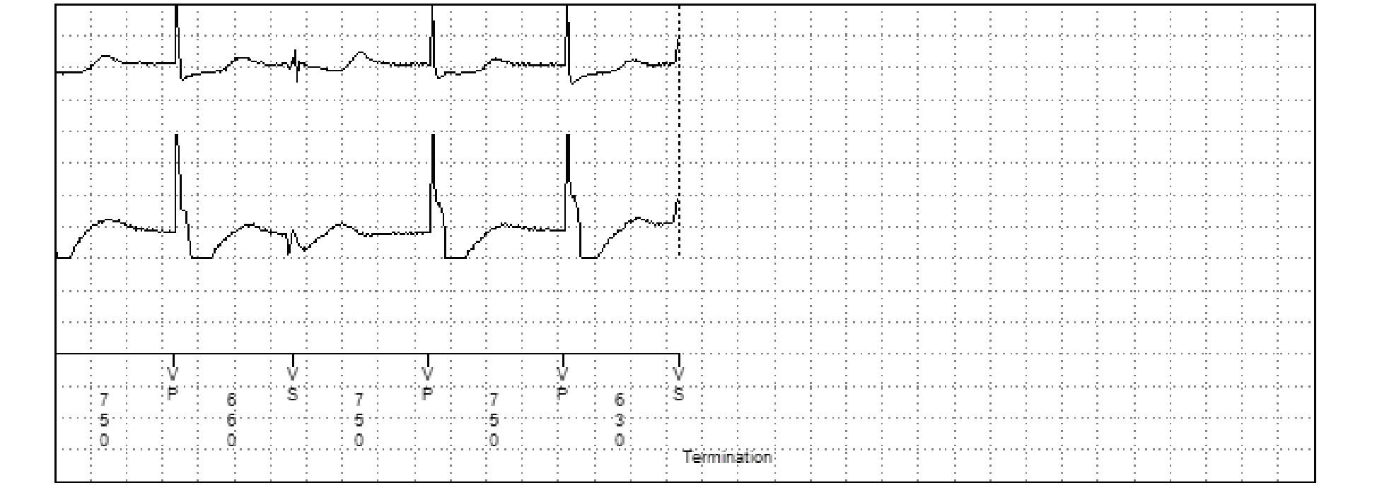
EGM

- What diagnosis is suggested by the interval plot? The plot shows an episode classified as VF, the duration of the ventricular cycles during the tachycardia being very variable; some cycles are very fast (150 ms) and others slower (500 ms); the 2 diagnoses suggested by this plot are: 1. a VF-type ventricular arrhythmia with intermittent undersensing (explaining the slower cycles); 2. oversensing of non-physiological signals (lead dysfunction, etc.).
- What type of arrhythmia is it? This is VF (very rapid, irregular, polymorphic arrhythmia); the shock channel (EGM2) allows the chaotic nature of the electrical activity to be clearly visualised.
- How do you explain this VS marker during initial detection? The cycle is classified as VS following ventricular undersensing of the previous complex.
- How is the initial detection counter programmed in the VF zone? The initial counter is set to 30/40; it is filled because 30 cycles are classified as either VF or FVT (FS and TF cycles fill the same VF counter, the FVT counter being set to via-VF) with only 3 cycles classified as VS (i.e. 30 rapid cycles out of 33); the cycles labelled FS and TF do not need to be consecutive; the existence of a few undersensed cycles does not prevent the counter from being filled and does not compromise patient safety.
- How do you explain these VS markers? Capacitor charging begins following the FD marker, a maximum output shock being the first therapy programmed for this type of arrhythmia; during capacitor charging, even if the ventricular cycles detected are very rapid and correspond to the VF zone, they are systematically labelled VS; in this example where sensing is imperfect, short and long cycles are all labelled VS during charging.
- How do you explain device function at the end of charging? At the end of charging, both fast and slow cycles are labelled VS; a shock is delivered if at least 2 out of 5 cycles are considered fast; here the first cycle following the CE marker is fast (280 ms), the second is slow (450 ms), the third is fast (190 ms) and so the shock is delivered (CD).
Points to remember
- One of the special features of MedtronicTM devices is that the counting method differs completely between the VT zone (consecutive cycles) and the VF zone (probabilistic counter).
- This diagram explains how the VF counter works; it is a probabilistic counter (X/Y rapid cycles) which requires a minimum of 75% rapid cycles (possible settings: 9/12, 12/16, 18/24, 24/32, 30/40, etc.).
- Ventricular fibrillation is by definition a rapid, chaotic arrhythmia, with ventricular signals of low and/or variable amplitude; all these characteristics increase the risk of undersensing, with the occurrence of signal amplitudes below the programmed sensitivity (nominal value of 0.3 mV) and often with great beat-to-beat variation in amplitude, which can fool the sensing circuit of the defibrillator because it uses a sensitivity level that adapts in relation to the previous signal.
- The value of 75% fast cycles was chosen to achieve an optimum balance between correct sensing of ventricular fibrillation (tolerating a certain number of pseudo-long cycles as a result undersensing) and the need to avoid filling counters in the presence of T wave, P wave or R wave oversensing (frequently associated with 50 % short cycles).
- This plot shows intermittent undersensing during VF but no significant impact on detection of the episode, with the probabilistic counter tolerating a maximum of 25% long cycles related to undersensing.
Practice out loud
Just describe the tracing. What do you think is going on? Describing the numbers can help.
00:00
0%
—
From Medtronic ICD — clinical cases by P. Bordachar, A. Thiyagarajah, L. Fontagne, M. Strik, S. Ploux. Published by Cardiocases. Every numbered marker on a recording is explained in the list beneath it; tap a marker on the trace or an entry in the list.
Order hardcopyon Amazon