
Chapter 2 · Therapies · Case 1
Episode of ventricular fibrillation and high defibrillation threshold
Patient and episode
Patient
- 57-year-old obese man with ischemic cardiomyopathy and ejection fraction of 25% implanted with an Atlas dual-chamber defibrillator for primary prevention; patient treated with amiodarone for paroxysmal AF; chest pain for 1 hour followed by prolonged syncope with, according to his wife, several jerks corresponding to electric shocks
Summary
- Episode of VF lasting more than one minute Six maximum electric shocks were delivered for this episode
The recording
Tap a number on the trace, or an entry in the list below
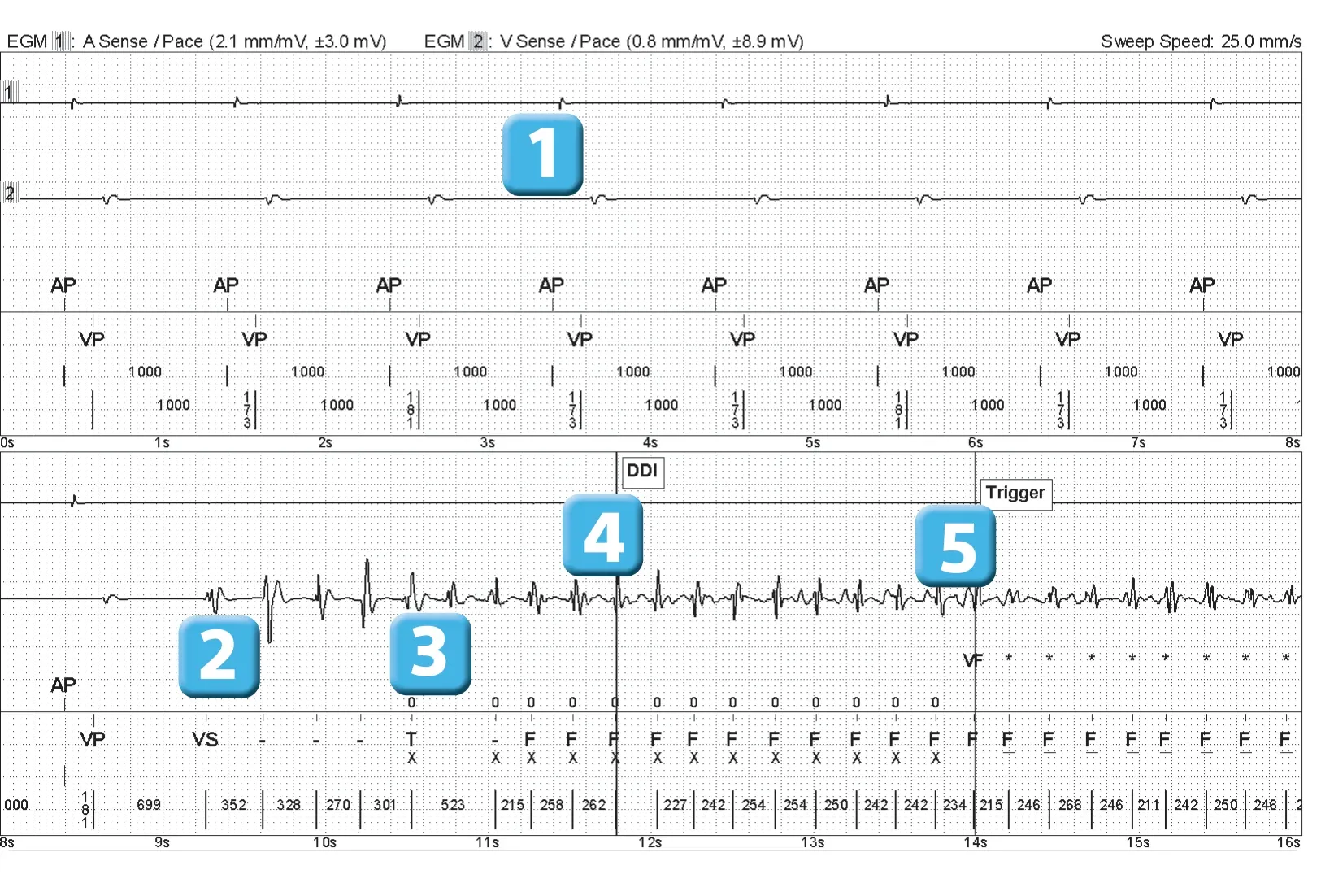
- rhythm stimulated rhythm in the atrium and ventricle (AP-VP) at minimum frequency (60 beats/minute)
- PVC
- Unclassified cycles followed by a classified cycle in the VT zone; note the subsequent under- sensing of a ventriculogram explaining the unclassified cycle that follows
- episode mode (DDI) after 4 cycles (3F + 1T) on this old platform
- episode of VF, triggering of EGM recording and start of capacitor charging (*)
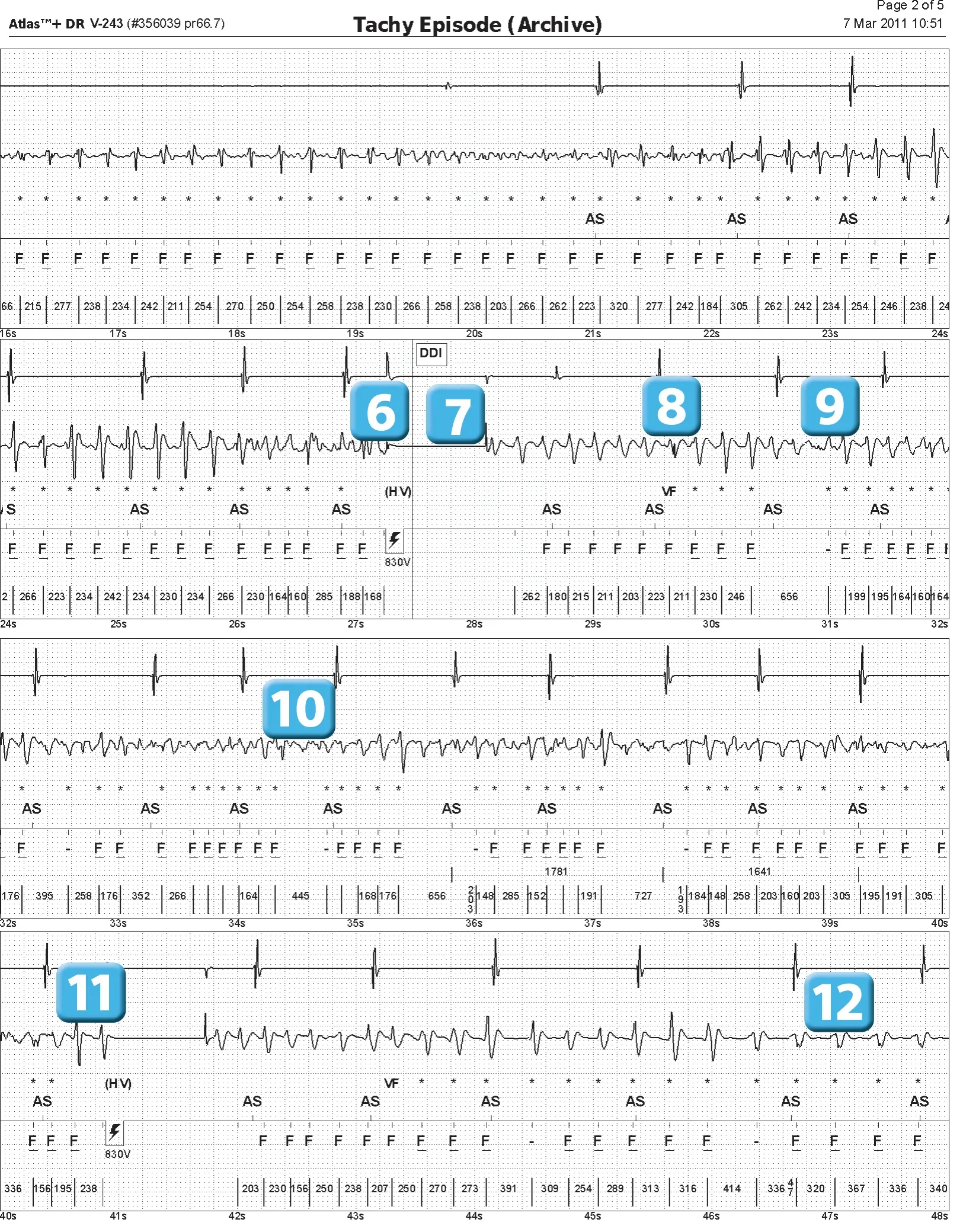
- 36 joules electric shock
- 1-second blanking post-shock
- shock failure, redetection of VF (after 6 F cycles) and new capacitor charging
- undersensing of low-amplitude ventricles
- new undersensing
- second shock of 36 joules
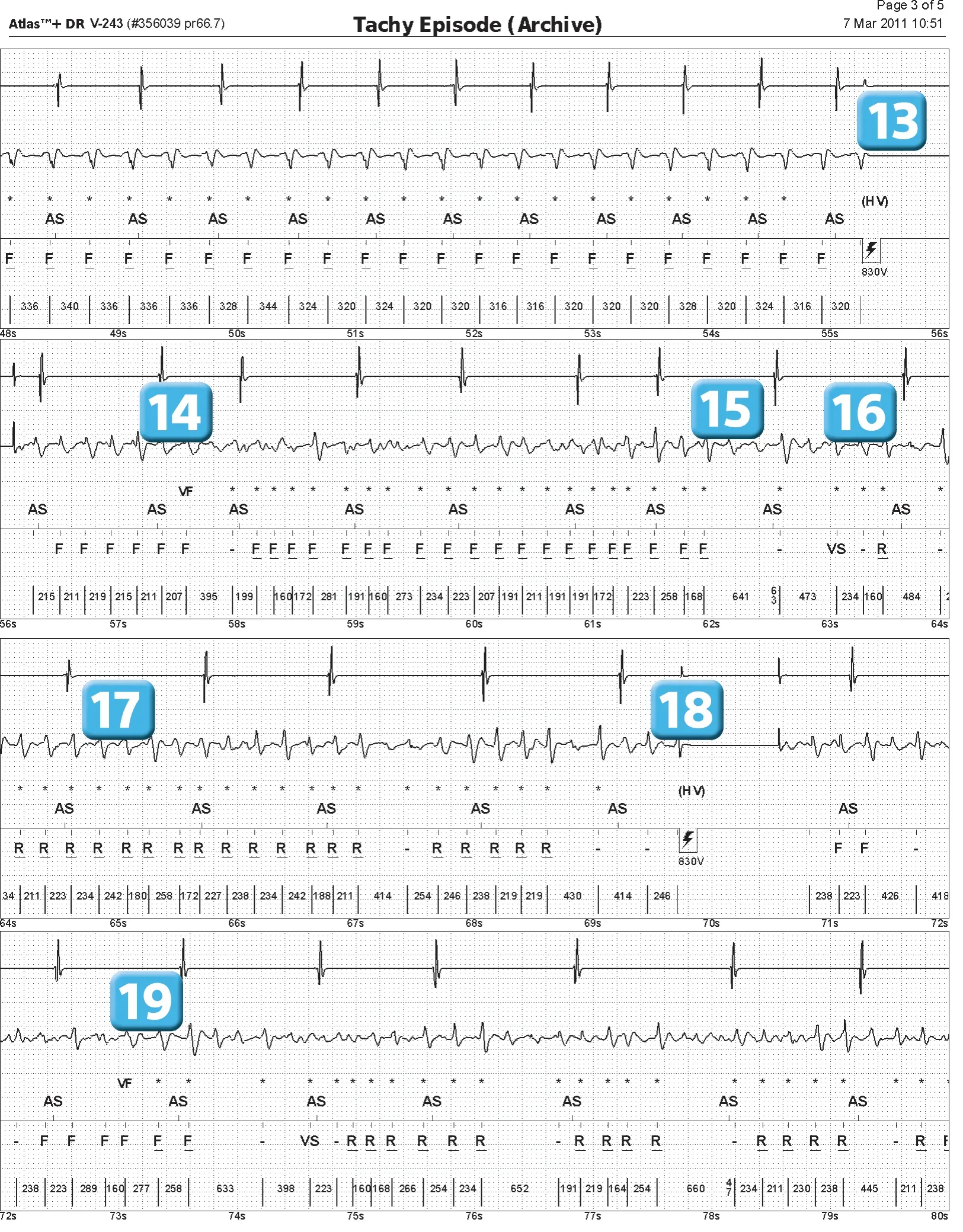
- shock failure, VF redetection and capacitor charging; arrhythmia stabilizes into organized VT
- third shock of 36 joules
- shock failure (deterioration to VF), redetection of VF and capacitor charging
- new undersensing marked
- false long cycle noted VS meaning that the current cycle and the average of the previous 4 cycles are outside the VT/VF zones
- Following this VS cycle, R markers were emphasized rather than F markers during charging.
- fourth shock of 36 joules
- shock failure, redetection of a VF and capacitor charging
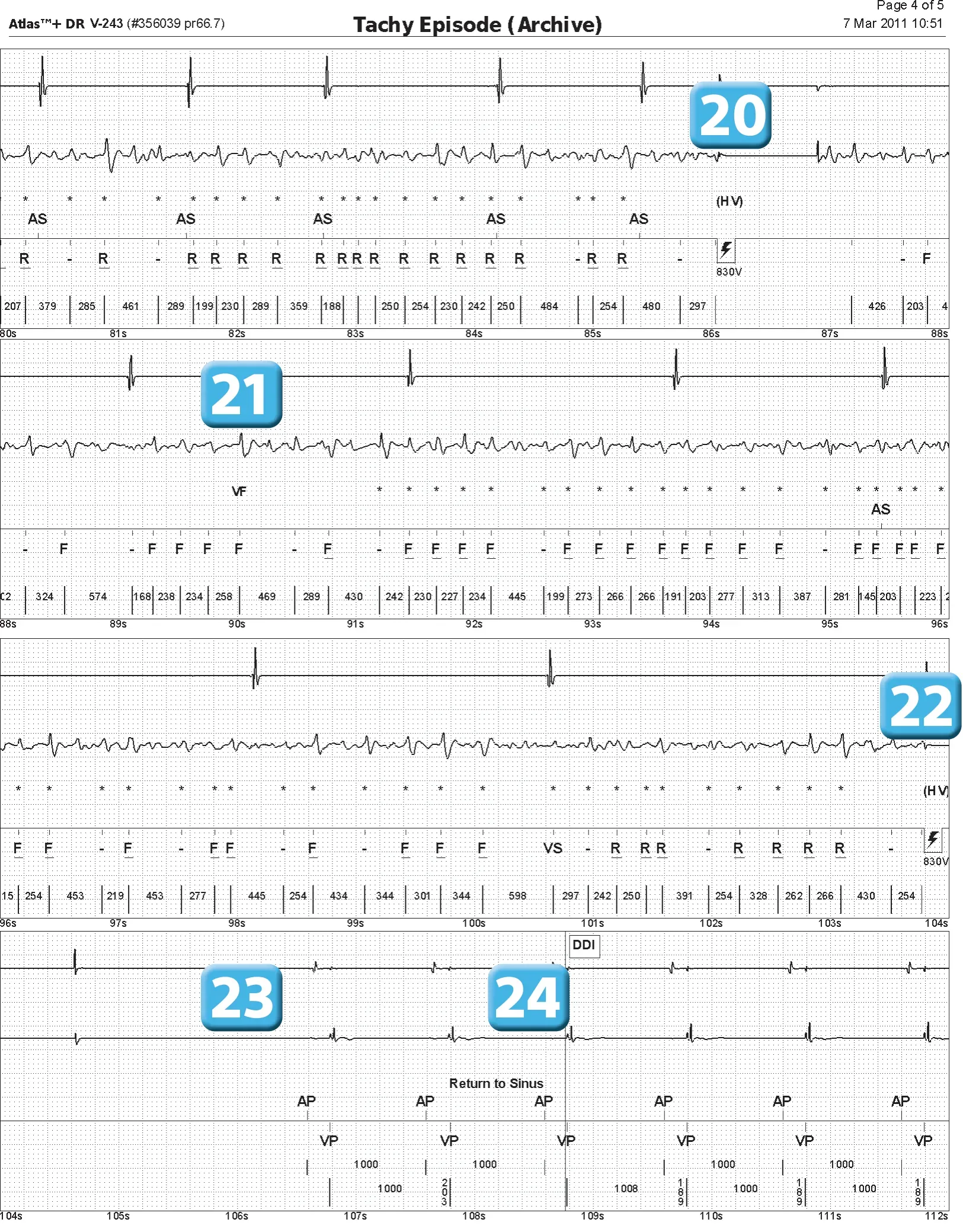
- Fifth shock of 36 joules
- shock failure, VF redetection and capacitor charging
- sixth shock of 36 joules
- following the shock, 1-second blanking phase and 2-second period without stimulation possible, explaining the pause
- shock effective and sinus return diagnosed (3 consecutive P waves)
Points to remember
- This trace was recorded on an Atlas defibrillator, which is an older model of defibrillator, explaining the differences in markers compared to more modern devices. Given their age, it is now highly unlikely that a device corresponding to this platform will be tested.
- Implantable defibrillators have historically been developed to prevent the risk of sudden death and reduce malignant ventricular arrhythmia by electric shock; in this patient, the arrhythmia is extremely rapid, polymorphic, and disorganized from the outset and corresponds to VF; Any attempt to reduce this type of arrhythmia through anti-tachycardia pacing seems doomed to failure, and electric shock remains the standard treatment in this context.
- For an Abbott defibrillator, a series of 6 electric shocks can be programmed in the VF zone; the probability of success of a shock after a maximum of 6 attempts is limited; conversely, it is preferable to limit the number of shocks if therapies are inappropriate.
- The arrhythmia episode presented by this patient is extremely concerning in that it was only reduced by the sixth shock at full power, the last available therapy. This tracing highlights the fact that the defibrillation threshold is not a fixed value; in this case, several shocks at maximum energy were ineffective, whereas the sixth shock of the same amplitude solved the very worrying situation.
- An electric shock is effective if a sufficient critical mass of myocardium is depolarized by establishing an intramyocardial voltage gradient. An electric shock is ineffective if a residual mass of fibrillating myocardium persists or if there is immediate reinduction in areas where the induced gradient is limited. When the first shock is ineffective, a vicious cycle ensues. Prolonged duration of VF increases hemodynamic deterioration, ischemia, and myocardial stretch, reducing the likelihood of success of the next shock.
- The effect of an electric shock varies depending on the energy delivered; for low energy, in the order of joules, an electric shock during a vulnerable period can induce arrhythmia; the upper vulnerability value corresponds to the lowest energy, applied during a vulnerable ventricular period, that does not cause ventricular fibrillation; this value is correlated with the defibrillation threshold; the probability of reduction then increases according to an exponential probability curve depending on the amplitude of the shock delivered ( synchronized with the R wave); above a certain value, the risk of reinducing arrhythmia also increases, limiting the chances of successful therapy; a shock with too high an amplitude can damage myocardial tissue
- in this patient, the third shock degraded a monomorphic and organized arrhythmia (the arrhythmia had stabilized) into a polymorphic and chaotic arrhythmia (new VF), which is typical in the presence of a high defibrillation threshold; the shock did not capture a sufficient amount of ventricular myocardium; on the contrary, it created sufficient pro-arrhythmogenic myocardial heterogeneity to generate multiple reentry circuits; it is therefore not surprising that the same amount of voltage delivered subsequently did not reduce the induced VF episode and that several shocks were necessary.
- In this patient, safety appears to be compromised; certain factors contributing to a high defibrillation threshold can be identified; some are difficult to modify: hypertrophic cardiomyopathy, obese patient; others are more accessible: treatment with amiodarone can be discontinued, which could lower the threshold, but could also increase the risk of ventricular arrhythmias ventricular
- The shocks delivered are biphasic, anodic polarity (anode in the ventricle, cathode at the device), with fixed tilt; the normal impedance of these shocks suggests that there is probably little hope of optimizing these parameters and programming a fixed pulse duration, for example.
- In this patient, a coil positioned in the coronary sinus was added; a shock delivered between two electrodes in contact with or in close proximity to the cardiac mass is more likely to be effective by extending the induced electric field over a larger volume.
Practice out loud
Just describe the tracing. What do you think is going on? Describing the numbers can help.
00:00
0%
—
From Implantable Cardioverter Defibrillator — clinical cases based on tracings by P. Bordachar, M. Strik, A. Thiyagarajah, S. Ploux. Published by Cardiocases. Every numbered marker on a recording is explained in the list beneath it; tap a marker on the trace or an entry in the list.