
Chapter 3 · Discrimination · Case 11
Dual-chamber discrimination and AF with aberrant conduction
Patient and episode
Patient
- 65-year-old male with severe ischemic cardiomyopathy; implanted with a dual-chamber defibrillator for primary prevention.
Summary
- Episode diagnosed in the VT zone
- A>V and rhythm ID discrimination
The recording
Tap a number on the trace, or an entry in the list below
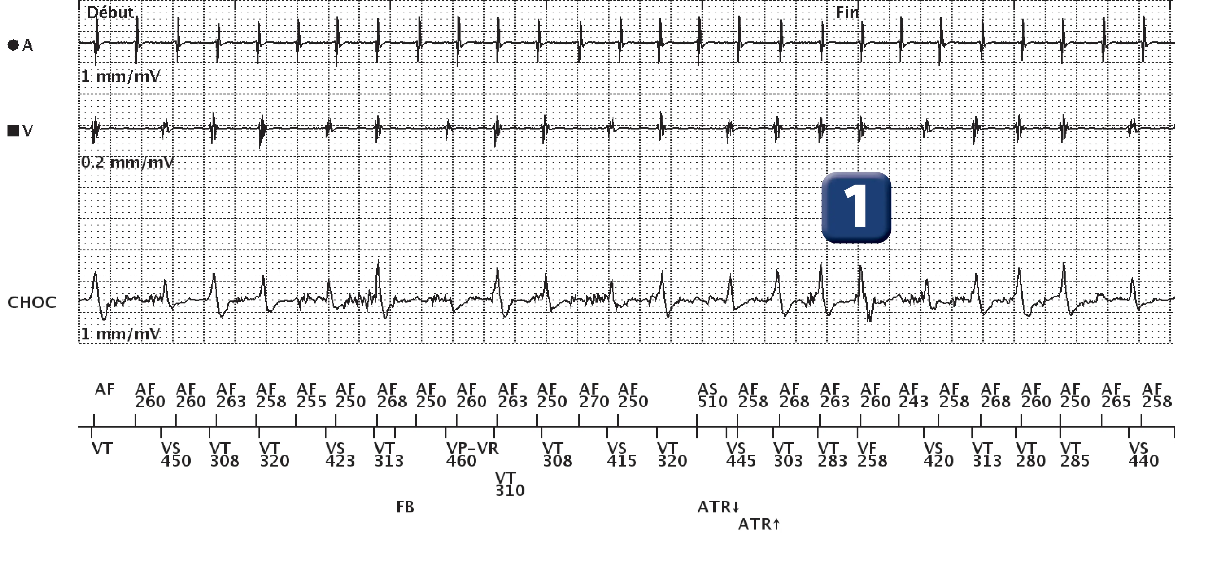
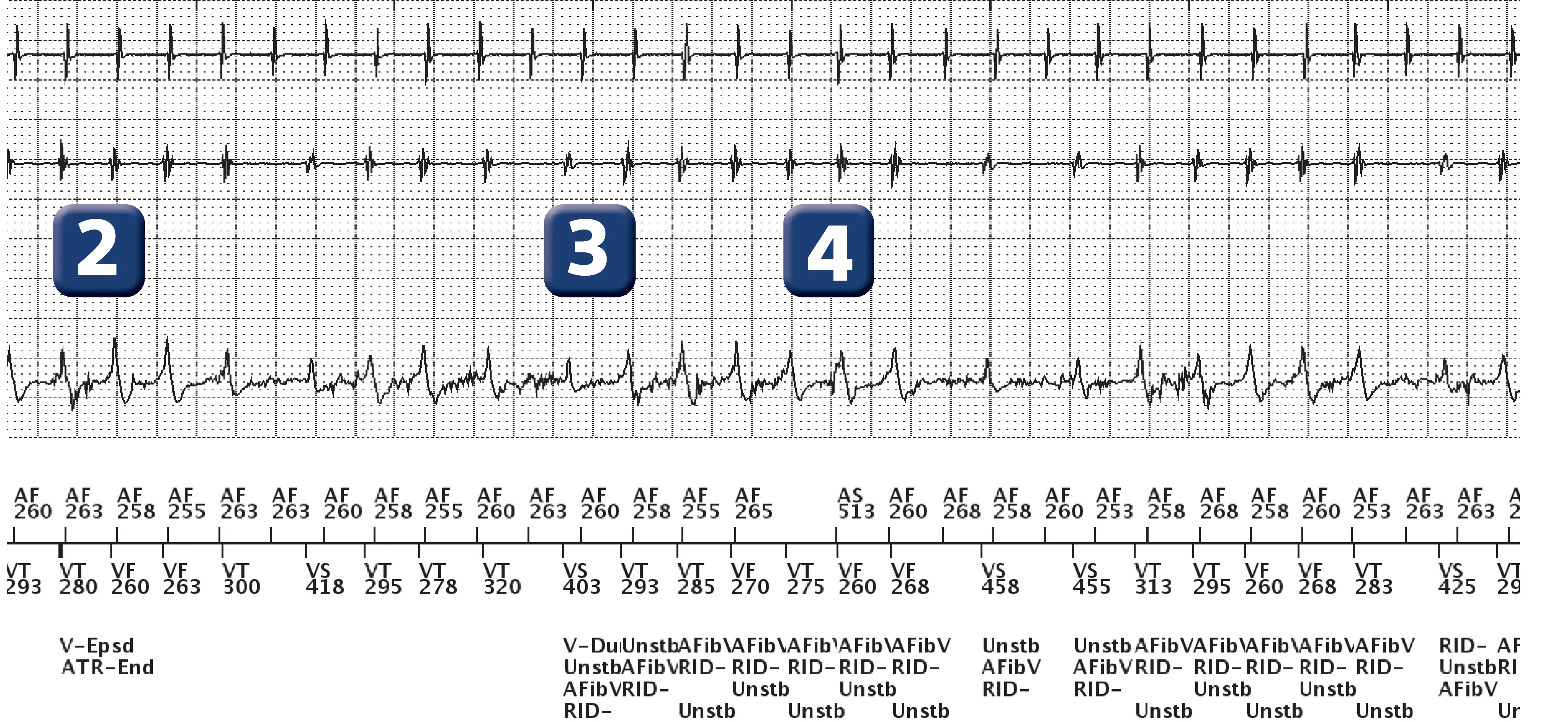
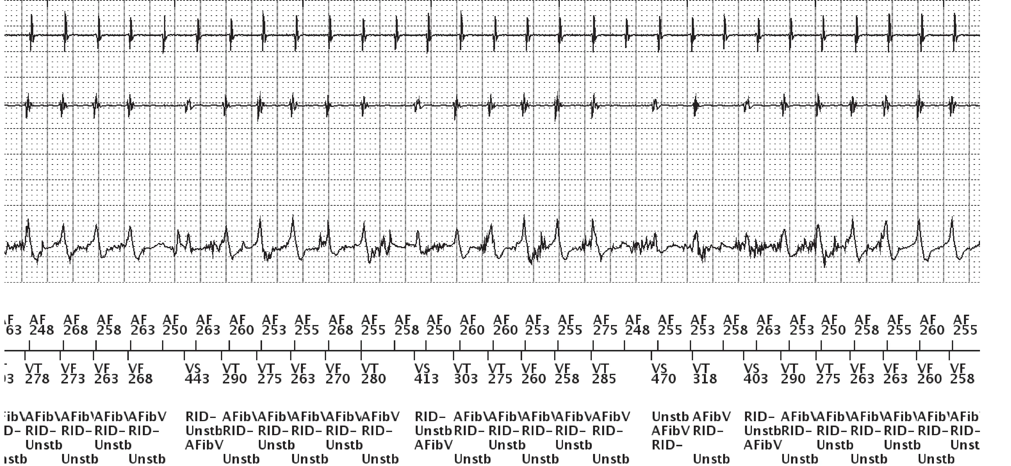
- Rapid atrial activity (probable atrial tachycardia) with rapid and variable atrio-ventricular conduction with alternating VS, VT then VF cycles
- Criterion 8/10 met (V-Epsd)
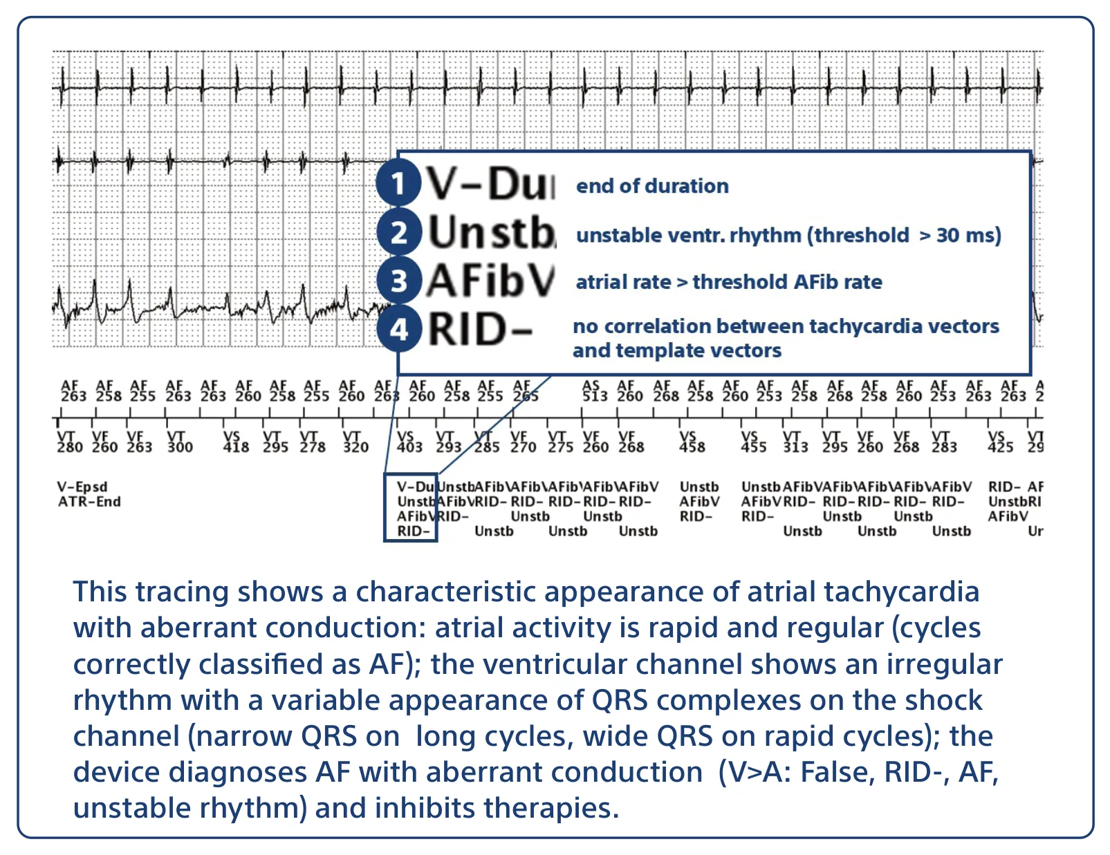
- At the end of the duration, decision not to treat in the presence of AF (atrial rate > A Fib rate threshold), unstable ventricular rhythm (stability value > programmed threshold of 30 ms) with absence of correlation between tachycardia vectors and reference vector (less than 3 cycles/10 correlated; note that cycles classified as VF are systematically considered uncorrelated); electrogram analysis shows variability in the appearance of ventricular electrograms on the shock channel, with widening on the shortest cycles
- Continuation of cycle-by-cycle analysis with no change in diagnosis (aberrantly conducted AF)
Points to remember
- Dual tachycardias and AF with aberrant conduction are probably the most difficult tachycardias to discriminate for a dual-chamber defibrillator; in fact, the V>A criterion does not allow a decision to be made, since in both cases the atrial rhythm is faster than the ventricular rhythm; in both cases, there is also no correlation between the vectors during tachycardia and the reference vector.
- When morphology analysis is combined with stability and the search for AF, stability determines the diagnosis when the vectors are not correlated and the atrial rate > A Fib rate threshold: if the rhythm is stable, the device diagnoses dual tachycardia and delivers therapies; if the rhythm is unstable, it diagnoses AF with aberrant conduction and inhibits therapies (as in this example)
Practice out loud
Just describe the tracing. What do you think is going on? Describing the numbers can help.
00:00
0%
—
From Boston Scientific ICD — clinical cases by P. Bordachar, M. Strik, A. Thiyagarajah, S. Ploux. Published by Cardiocases. Every numbered marker on a recording is explained in the list beneath it; tap a marker on the trace or an entry in the list.
Order hardcopyon Amazon