
Chapter 3 · Discrimination · Case 12
Dual-chamber discrimination and discrimination error due to signal variability on the shock channel
Patient and episode
Patient
- 49-year-old man; hypertrophic cardiomyopathy; implanted with an Autogen dual-chamber defibrillator
Summary
- Tachycardia episode with atrial rate equal to ventricular rate (V>A False)
- Initially, correlation of Rhythm ID: True; inhibition of therapies
- In a second step, Rhythm ID: False; AF: true + stable rhythm (threshold value set at 20 ms)
- All VT zone therapies are delivered (ATP + 6 shocks)
The recording
Tap a number on the trace, or an entry in the list below
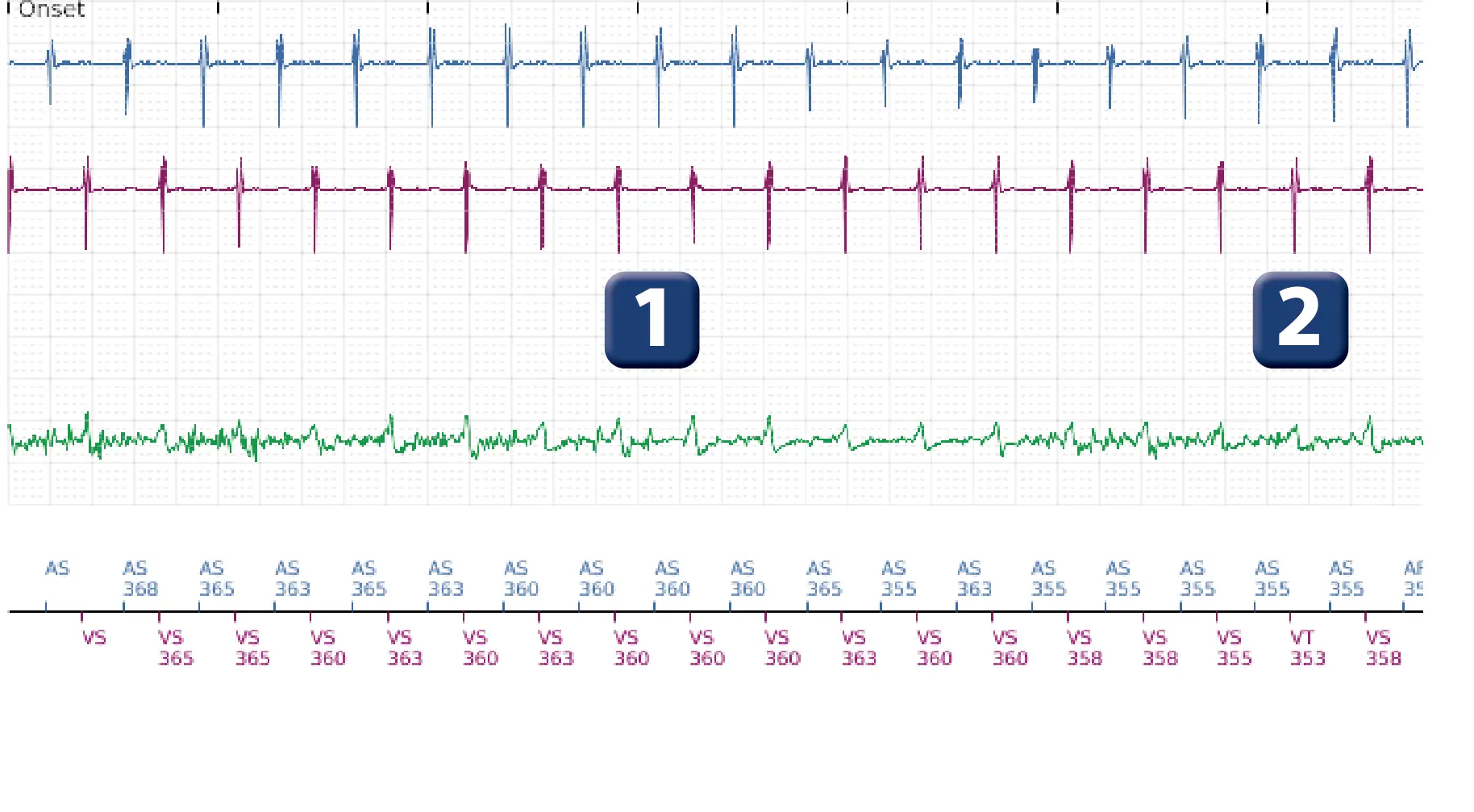
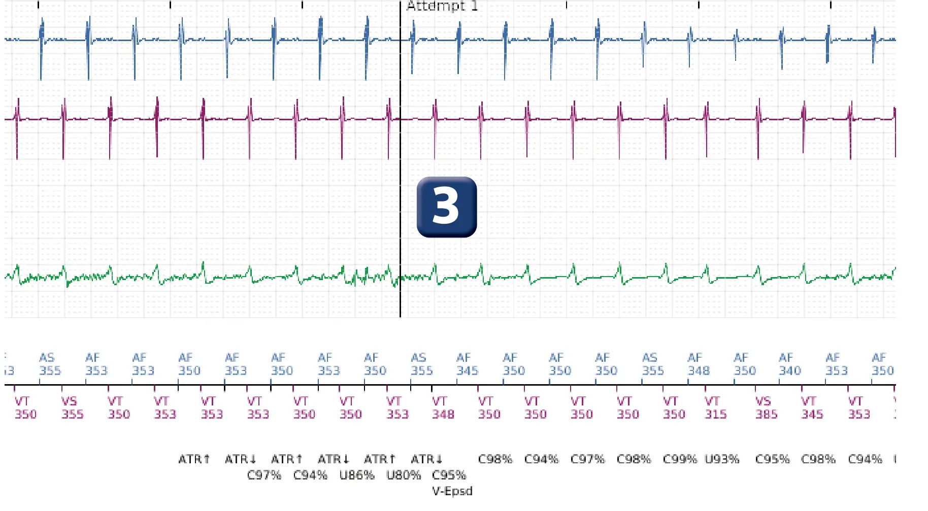
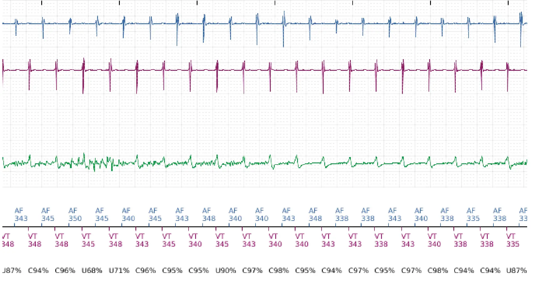
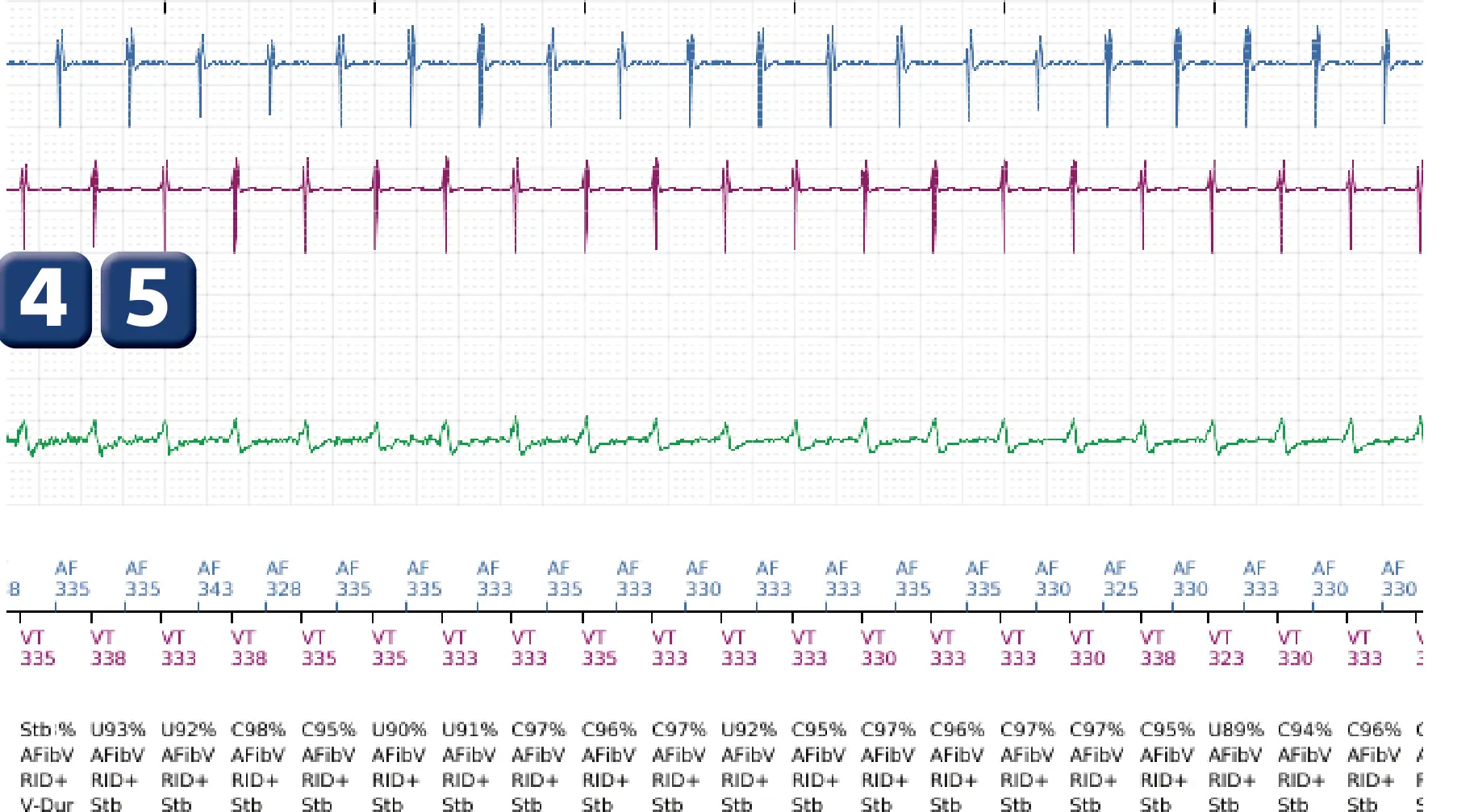
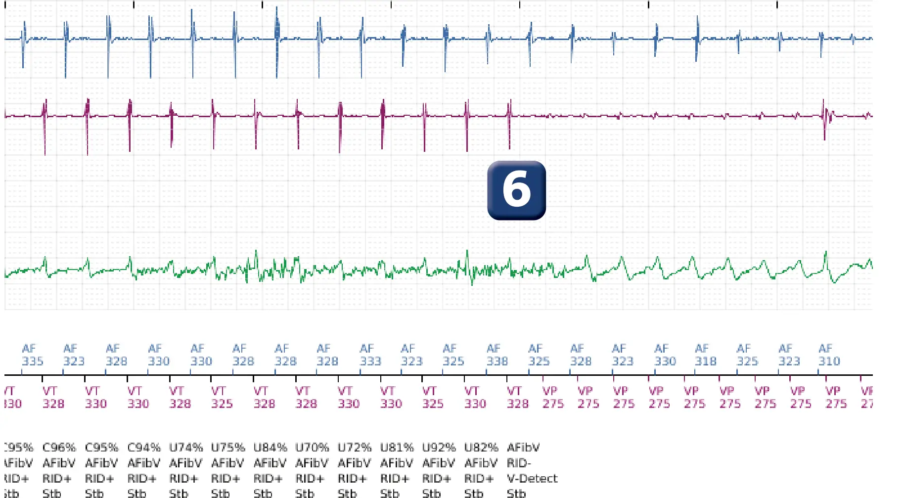
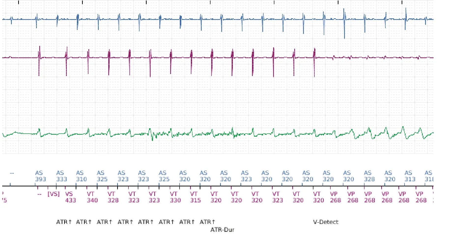
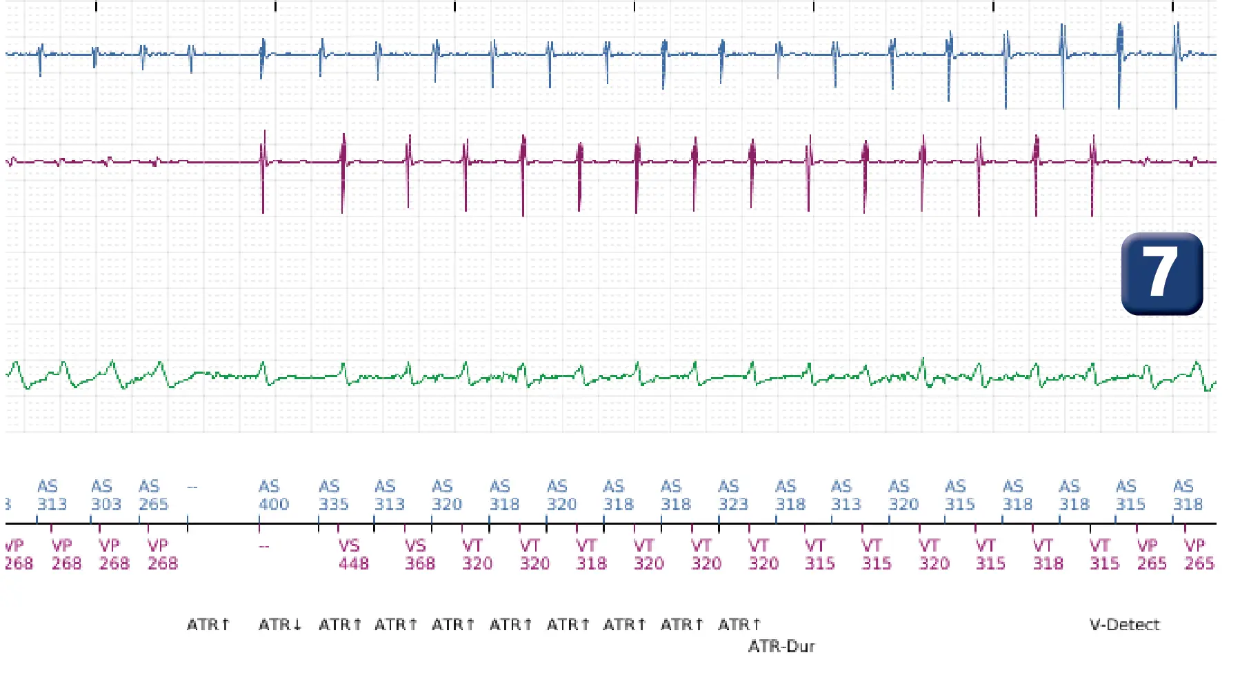
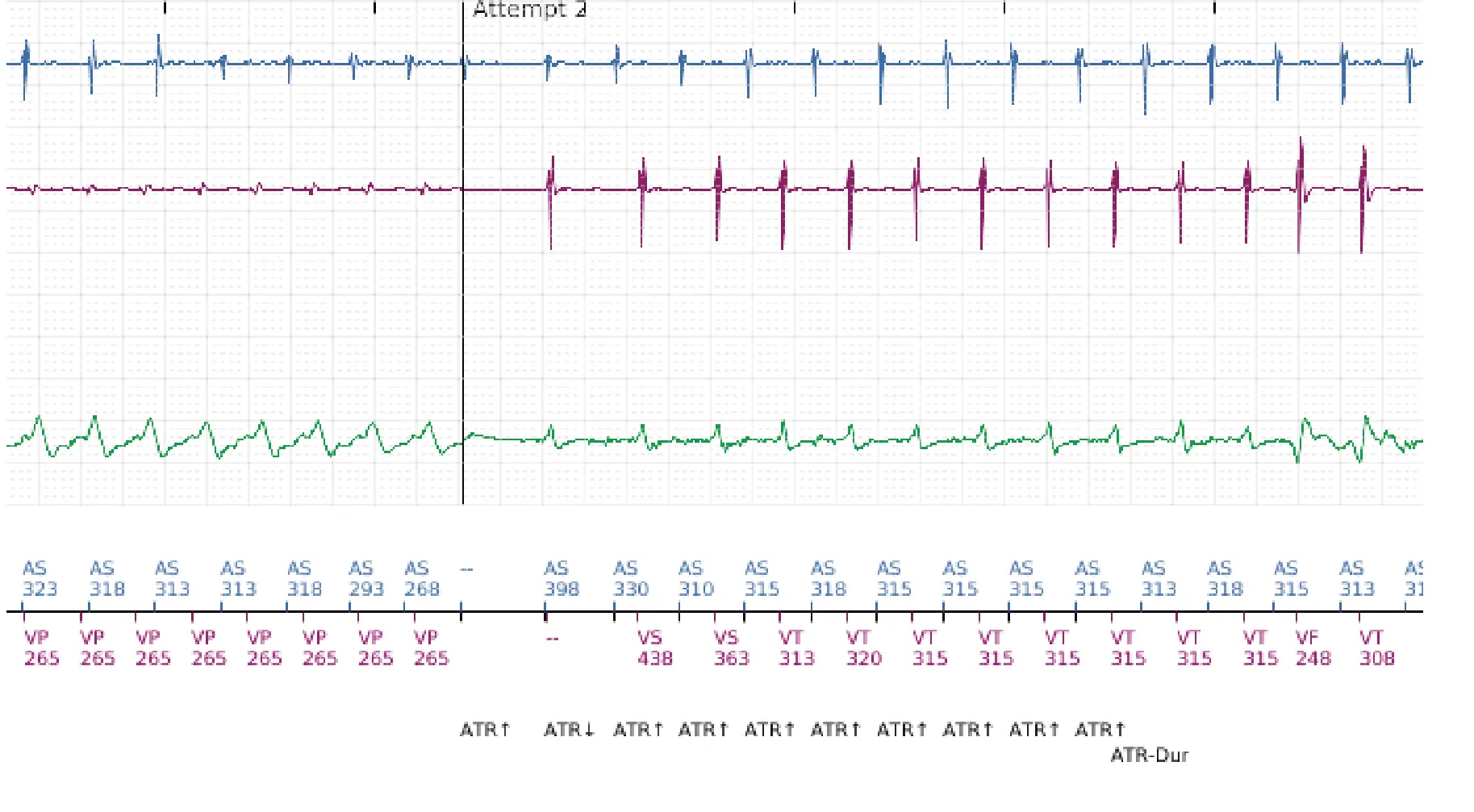
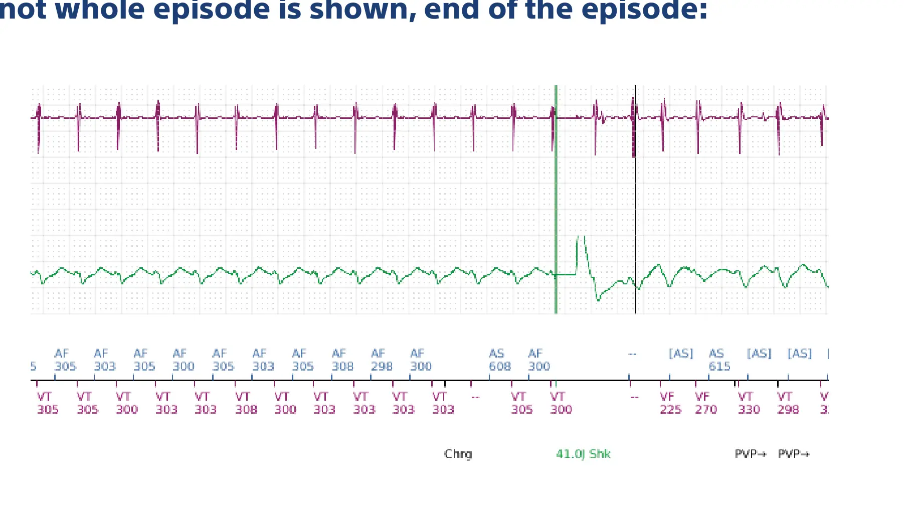
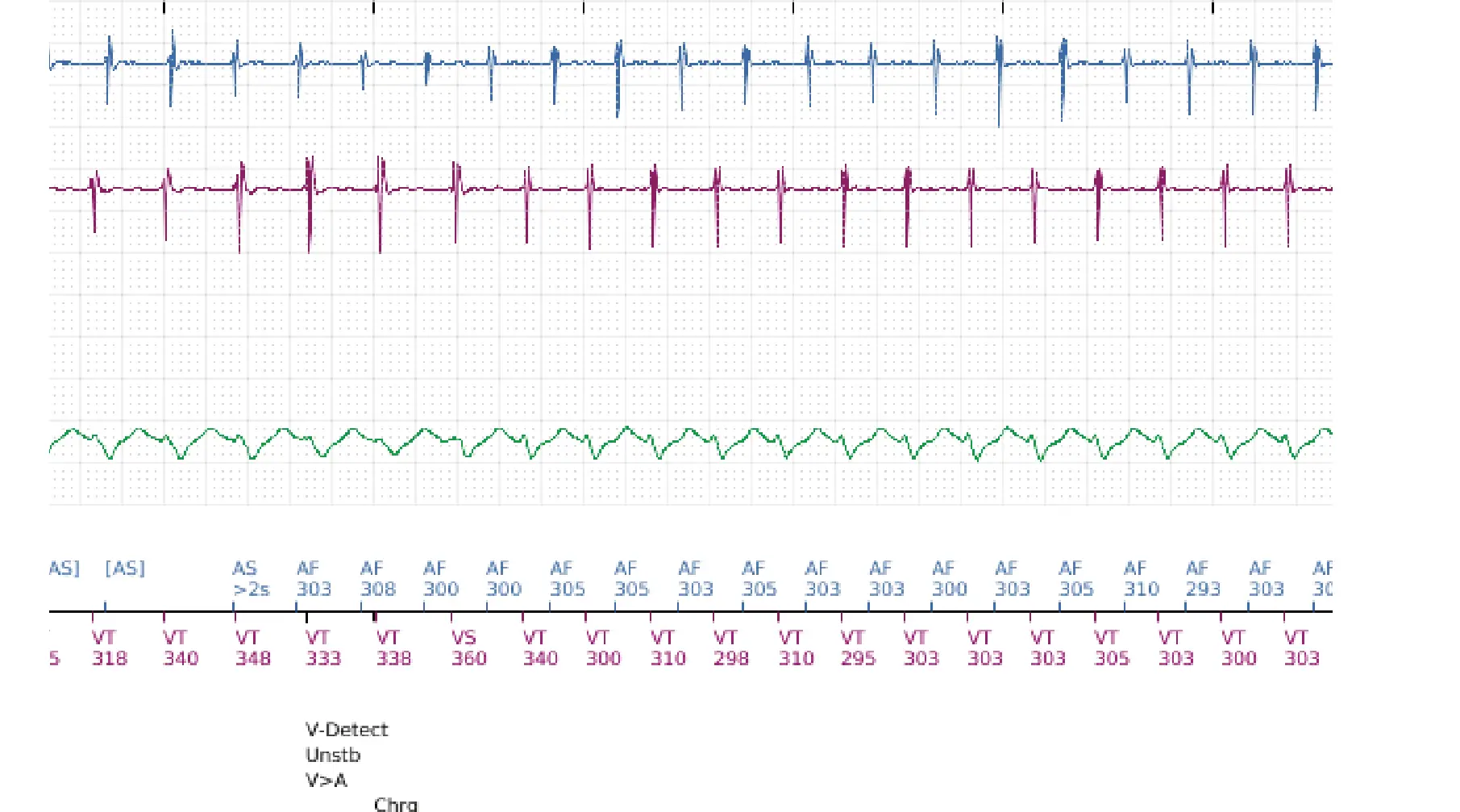
- Regular tachycardia 1/1 (AS-VS)
- Acceleration of atrial cycles (cycles classified as AF) and ventricular cycles (cycles classified as VT)
- After 8 consecutive VT cycles, V-Epsd marker; signal quality in the shock channel is variable (some cycles are considered correlated, others uncorrelated)
- End of duration; for the device, the rhythm is stable with a diagnosis of AF; therapies are not delivered because RID+ (at least 3 correlated ventricular complexes out of 10).
- Cycle-by-cycle discrimination continues, still with RID+.
- Signal variability on the shock channel; RID- (less than 3 correlated cycles out of the last 10); stable rhythm with diagnosis of AF: suspicion of double tachycardia and first therapy is delivered.
- Redetection and new therapies; all therapies in the zone are then delivered
Points to remember
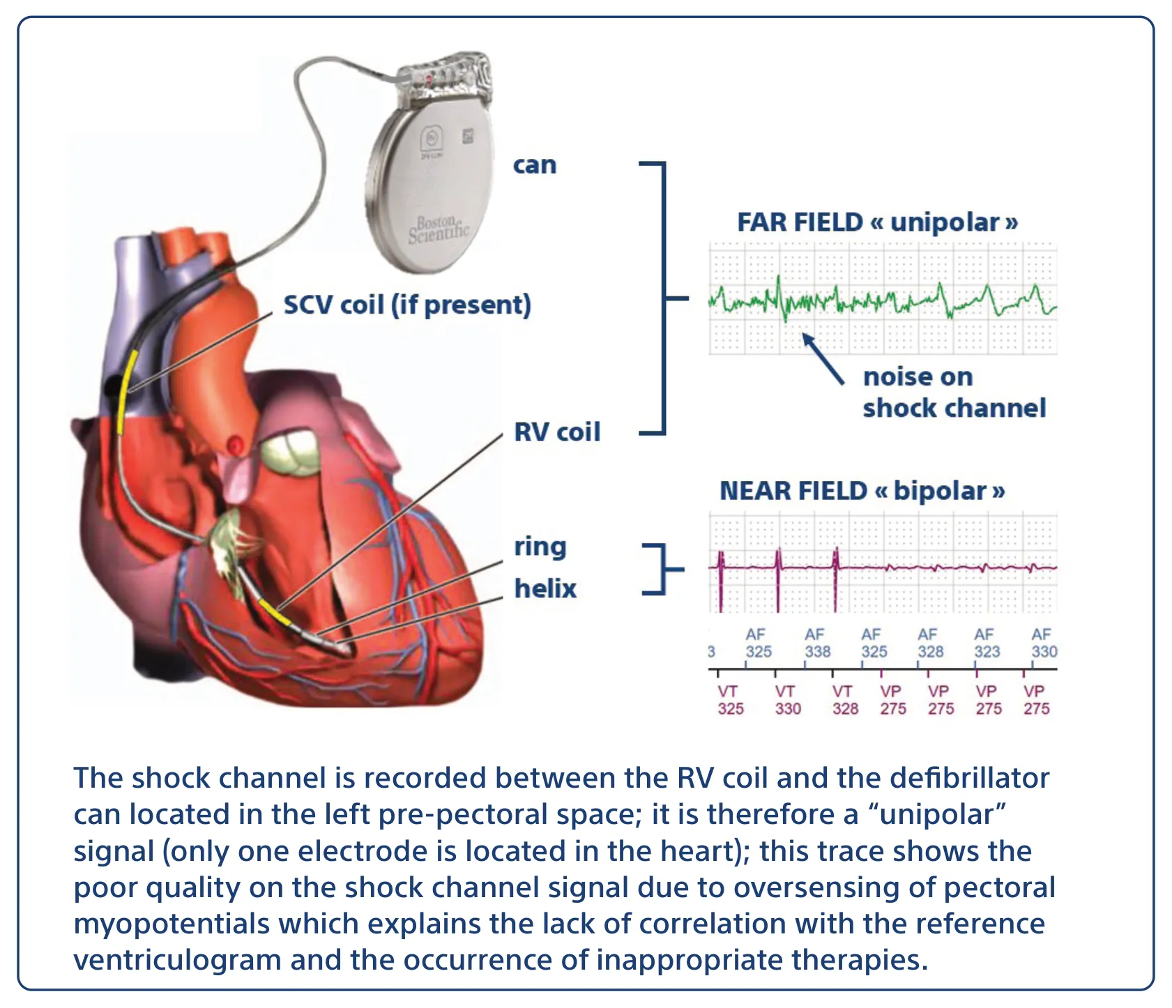
- This trace shows an example of inappropriate therapies for supraventricular tachycardia; the ventricular rate is not higher than the atrial rate (V>A rate: false); initially, discrimination is correct with ventricular cycles correlated with the reference ventricular electrogram (RID+); in a second phase, a series of inappropriate therapies is delivered following variable signal quality on the shock channel (RID- and stable rhythm)
- In this example, signal quality was impaired by the oversensing of myopotentials on the shock channel.
Practice out loud
Just describe the tracing. What do you think is going on? Describing the numbers can help.
00:00
0%
—
From Boston Scientific ICD — clinical cases by P. Bordachar, M. Strik, A. Thiyagarajah, S. Ploux. Published by Cardiocases. Every numbered marker on a recording is explained in the list beneath it; tap a marker on the trace or an entry in the list.
Order hardcopyon Amazon