
Chapter 4 · Discrimination · Case 6
Limitations of discrimination based on Wavelet
Patient and episode
Patient
- Male implanted with a single-chamber defibrillator (Evera XT VR) for ischaemic cardiomyopathy, hospitalised for syncope with a shock from his device.
The recording
Tap a number on the trace, or an entry in the list below
Rate / interval plot
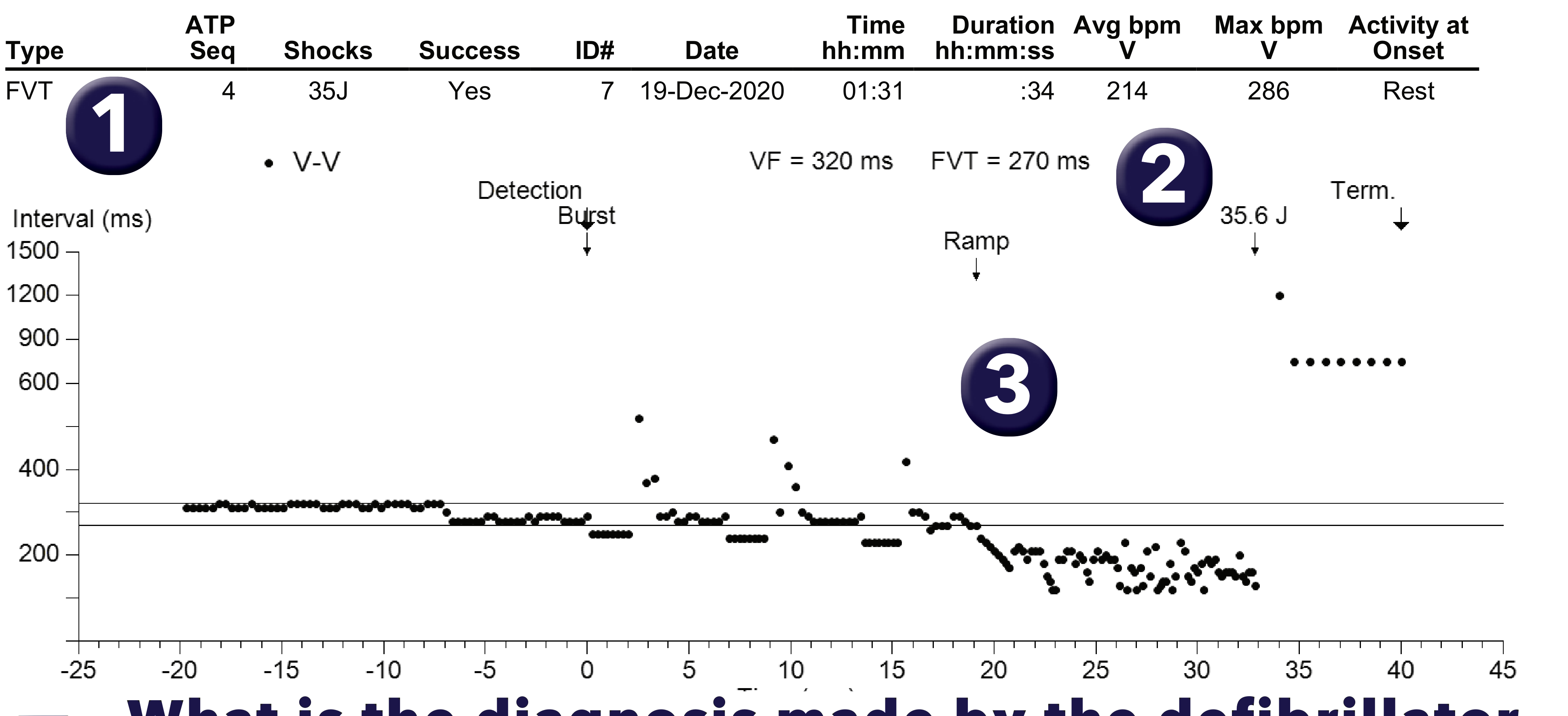
From the interrogation
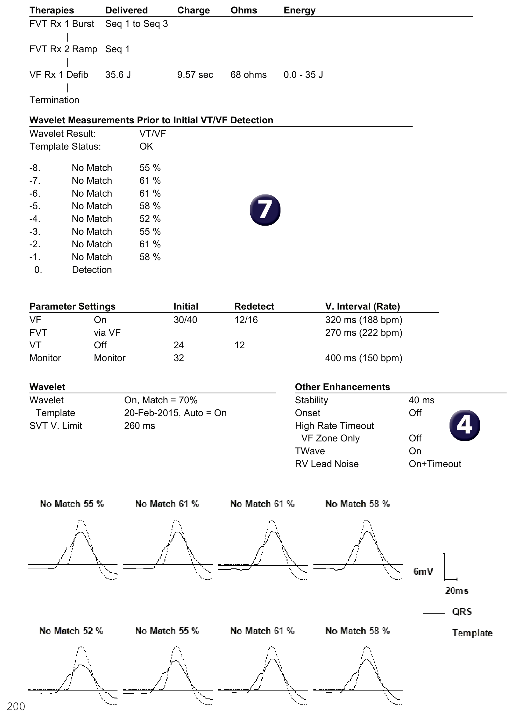
EGM


- What is the diagnosis made by the defibrillator for this episode? This episode has been classified FVT by the device.
- How many detection zones are programmed? Two detection zones have been programmed.
- What diagnosis does the interval plot suggest? The graph shows a rhythm initially detected in the FVT zone with 3 bursts, then a ramp which appears to accelerate the tachycardia into the VF zone; a shock re-establishes a viable rhythm.
- How many discriminators are programmed? Wavelet and Stability are programmed.
- What does the WV marker mean? Initially, the patient presented with a regular, monomorphic tachycardia; the device diagnosed SVT based on Wavelet (WV); no therapy was delivered.
- How do you explain that a burst was delivered? The VF counter was completed again after 30 cycles classified as FS; the appearance of the ventriculograms appeared more or less identical to that observed at the start of the trace, but Wavelet concluded VT and therefore did not withhold therapies.
- What do you think of the Wavelet results? The similarity percentages ranged from 52% to 61% for the 8 complexes analysed and were therefore below the programmed similarity threshold (70%), which explains the diagnosis of VT/VF.
- What effect does the ramp have? The ramp accelerates the tachycardia into a very rapid VF.
- What is the effect of the shock? The shock terminates the arrhythmia.
Points to remember
- This patient had multiple episodes of atrial tachycardia which were well discriminated on the basis of morphology; he also had a few episodes which were poorly discriminated with inappropriate therapies; in this example, the discrimination error resulted in the occurrence of a ramp which induced a potentially lethal VF; a shock re-established a viable rhythm.
- The specificity of discrimination based exclusively on Wavelet is imperfect in certain patients; discrimination only applies to initial detection and an error may lead to multiple inappropriate therapies (no discrimination if tachycardia continues after the first therapy delivered).
- Certain factors may explain the imperfect specificity in certain patients:
- Wavelet performance depends directly on the quality of the discrimination signal (EGM2), which is usually the Can-RVCoil channel. The richer the signal, the greater its amplitude. During implantation, therefore, care must be taken to ensure that the Can-RVCoil signal amplitude is >3mV. This amplitude can be measured using the Medtronic analyzer, by collecting the unipolar signal from the RV coil and measuring the peak-to-peak amplitude (to do this, print out the signal).
- Outside the implantation procedure, you can measure the amplitude of the Can-RVCoil signal by selecting this channel on the programmer and making a printout. If the amplitude is too low, a different analysis vector can be selected (source EGM2: Can-RingRV; TipRV-CoilRV; TipRV-RingRV; Can-SCV; Can -RV-SCV). Note that changing the EGM2 vector deactivates the noise algorithm (which only works with the Can-CoilRV vector). It should also be borne in mind that near-field signals (TipRV-CoilRV; TipRV-RingRV) are generally less rich and therefore less effective for morphology analysis.
- On the other hand, the signal amplitude may be too large for the EGM2’s sampling range (nominal +/-12mV), which may result in signal amputation. In this case, you may need to select a larger scale (Parameters->Data collection configuration->EGM2 range).
- Automatic reference collection does not include frequency or AV delay hysteresis, so it cannot operate on a CRT-D. On a CRT-D, we recommend acquiring a Wavelet reference manually at each consultation.
- The Can - RV Coil signal can be acquired using the analyzer by connecting the unipolar signal on the RV Coil electrode and by measuring the peak-to-peak amplitude.
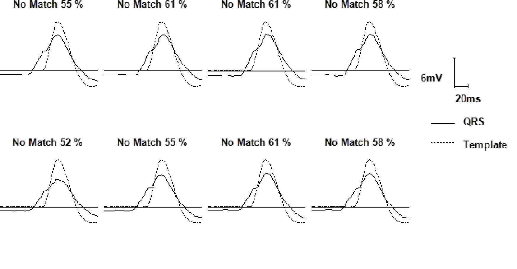
Practice out loud
Just describe the tracing. What do you think is going on? Describing the numbers can help.
00:00
0%
—
From Medtronic ICD — clinical cases by P. Bordachar, A. Thiyagarajah, L. Fontagne, M. Strik, S. Ploux. Published by Cardiocases. Every numbered marker on a recording is explained in the list beneath it; tap a marker on the trace or an entry in the list.
Order hardcopyon Amazon