Discrimination error in a triple-chamber defibrillator
Tracing
Manufacturer Boston Scientific
Device CRT
Field Management of atrial arrhythmias
N° 5
Patient
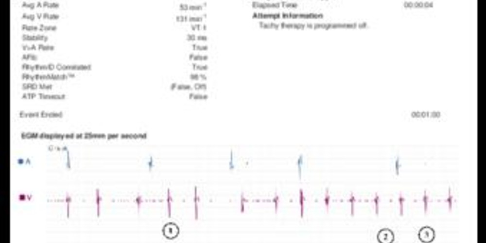
A 60-year old woman with a history of ischemic cardiomyopathy was implanted with a triple-chamber INCEPTA CRT-D. Interrogation revealed a VT episode.
Graph and trace
- probably sinoatrial activity with dissociated fast ventricular rhythm. Certain ventricular activations are classified as premature contractions (PVC), others as conducted (VS).
- VT episode with atrio-ventricular dissociation.
- wrongful analysis of morphology with high correlation percentages when compared with the template (>94%).
- despite the error in the morphology analysis, a VT is diagnosed since V>A. No therapies were programmed in this zone (ATP disabled).
- continuation of the tachycardia without delivered therapies.
Other articles that may be of interest to you

This EGM illustrates certain difficulties of discrimination in a triple-chamber device and the limits of morphology analysis. Two different approaches are programmable in the defibrillators of this manufacturer: an analysis based on parameters of stability and sudden onset and an analysis based on evaluation of ventriculogram morphology. The latter option allows for combined parameter programming depending on the presence and proper function of an atrial lead: threshold of AF rate, rate V > rate A, stability and morphology correlation. These options are only available for initial detection. When rate V > rate A is enabled and the tachycardia meets this criterion, this parameter has the priority over all other discriminating parameters like shown in this example where VT diagnosis occurred despite an error in morphology analysis.
For correct morphology analysis, acquiring a reference morphology (the template), against which the ventriculograms during tachycardia are compared, is an essential step of the process. Two automatic methods are available for programming: ‘passive’ or ‘active’. When ‘passive’ is programmed, the device searches for intrinsic ventricular activity every 2 hours. In this patient, the ‘passive’ option was selected. During the active method, the device searches for a template every 28 hours by temporarily modifying stimulation parameters (favoring intrinsic ventricular conduction, decreasing lower rate, and elongating the AV delay). The active method is the method of choice for patients who have high cumulative percentage of ventricular stimulation (in totally dependent patients, discrimination should be turned off), which is the case for most resynchronized patients. In addition, it is possible to record a template in real-time using the programmer. In this example, it seems obvious that the automatic acquisition of the template was done during tachycardia which explains the error of morphology analysis.