T wave oversensing

Tracing
N° 24
Manufacturer Biotronik Device PM Field Sensing
Patient

71-year-old man, implanted with a Biotronik Evia DR-T dual-chamber pacemaker for syncopal complete atrioventricular block; routine control; numerous PVCs recorded in the device's memory.
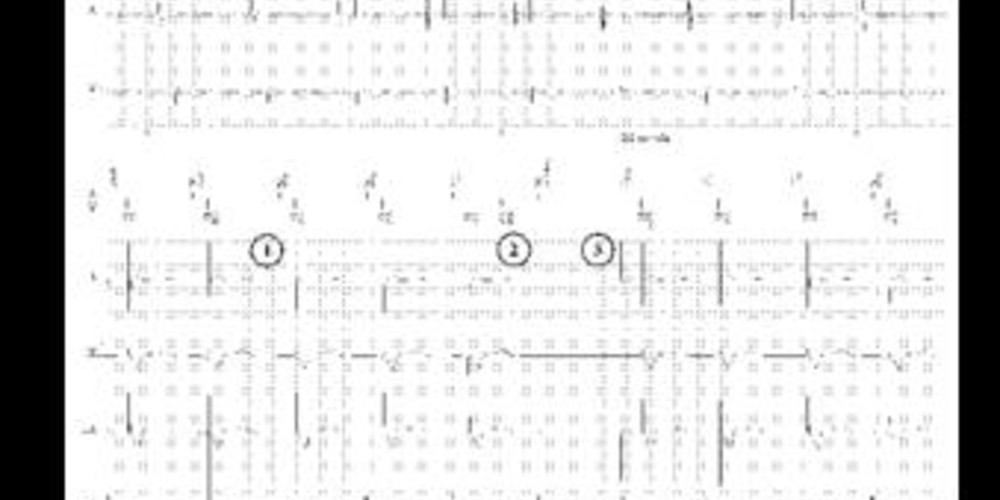
Graph and trace
  1. atrial sensing and ventricular pacing;
  2. ventricular pacing and T wave oversensing; sinus P wave sensed in the PVARP (prolongation of the post-PVC PVARP);
  3. atrial pacing and ventricular pacing.
Comments

The quality of the sensing of a signal is dependent on a certain number of its physical characteristics:

  1. its frequency spectrum: the frequency of a signal is expressed in hertz (Hz), and is the inverse of its period. A pacemaker amplifies incoming signals in a frequency range, on average, between 10 and 70 Hz corresponding to the cardiac depolarization signals. The signals located below and above this zone are filtered and are therefore rendered less or non-detectable by the system. In the ventricular channel, the R waves have a frequency spectrum between 10 and 30 Hz and are amplified. On the other hand, the T waves whose spectrum is less than 5 Hz are filtered. Similarly, signals of atrial origin collected in the ventricle usually have a very low frequency, and are most often filtered. The frequency spectrum of signals of muscular origin, such as the pectoral muscle (myopotentials), is superimposed on that of P waves and R waves. In the unipolar configuration, the sensing field extends from the distal electrode of the pacing lead to the pulse generator which is placed on or under the pectoral muscle with an increased risk of interference by the sensing of signals of muscular origin during certain physical exertions;
  2. its slope: this parameter describes the amplitude variation of the cardiac signal as a function of time, expressed in mV/ms. The pacemaker senses the fastest portion of the signal corresponding to the passage of the depolarization wave facing the electrode. If the depolarization signal is fragmented as is sometimes the case for an extrasystole, the slopes of its various components are often slower, with an increased risk of undersensing. At implantation, the pacing leads should ideally be positioned at a site where the depolarization slope is at least 1mV/ms in the ventricle and at least 0.5mV/ms in the atrium. The measurement of the slope of the signal is contingent on its processing, and in particular on the filters used. The latter differ depending on the measuring system, the external device for determining the thresholds or the prosthesis that will be permanently connected. The observed differences can be very significant. However, direct recording of the signal at implantation may be useful in searching for the site that yields the largest intrinsic deflection. The intrinsic deflection of an endocardial signal almost never occurs at the beginning of the corresponding signal on the surface ECG. For example, sensing of ventricular depolarization in a patient with complete right bundle branch block is very late. Similarly in the atrium, atrial sensing may occur at the end of the P wave of the surface ECG;
  3. its amplitude: the amplitude of the signal measured by the pacemaker corresponds to the amplitude of the signal which remains detectable by the pacing system after analysis of its frequency and the determination of the slope. It is expressed in mV. This is the parameter used at the end of the signal processing chain to determine the sensitivity level of the system. At implantation of a pacing lead, attention must be given to obtaining a signal corresponding to the band pass of the pacemaker, the intrinsic deflection of which is the fastest possible and of high amplitude. Amplitude levels of at least 5 mV in the ventricle and at least 2 mV in the atrium are typically targeted.

This tracing reveals an oversensing of the T wave, the ventricular channel being too sensitive. Automatic and adaptive cycle-to-cycle sensing, which is absolutely essential for the proper functioning of a defibrillator, is now available in pacemaker platforms. Pacemaker constraints in terms of sensing of fast low-amplitude signals are less than those of a defibrillator (necessity to sense and treat ventricular fibrillation). On the other hand, the oversensing of a cardiac (T wave, P wave) or extracardiac (myopotential, interference, etc) signal can be extremely problematic in pacemaker-dependent patients. In the presence of T wave oversensing, as observed in this patient, programming of a fixed rather than automatic sensitivity may be preferred.

Other articles that may be of interest to you