Single chamber Rhythm ID discrimination: VT
Tracing
Manufacturer Boston Scientific
Device ICD
Field Discrimination
N° 1
Patient
This 71-year-old man underwent implantation of an Incepta single chamber defibrillator for primary prevention in the context of ischemic cardiomyopathy. He was seen for a routine ambulatory visit.
Summary
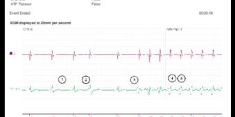
Episode diagnosed as VT at a rate of approximately 160 bpm; at the onset of the episode, the detection was based on Rhythm ID and the episode was treated with a burst of ATP, in absence of a correlation between the morphology of the ventricular EGM during tachycardia and the reference morphology (Rhythm ID correlated: False).

Graph and trace
Tracing
- spontaneous rhythm at 80 bpm;
- ventricular PVC;
- sudden onset of a relatively stable tachycardia stable with a distinctly different morphology on the high-voltage channel compared with the ventricular EGM during sinus rhythm;
- at the 3rd consecutive cycle in the VT zone, an episode is recorded in the memories;
- onset of the vector analysis (morphology and EGM synchronization) of the signal associated with the cycle following the third short cycle and comparison with the reference template;
- after 8 cycles in sinus rhythm, episode of VT (V-Epsd);
- Duration programmed at 2 sec, after which the discrimination (RID-) is in favor of VT (V-Detect); hence, for <3 out of 10 cycles (rolling window), the vector was correlated with the reference vector; à decision to treat;
- burst of ATP;
- successful burst and termination of the arrhythmia.
Other articles that may be of interest to you

EGM recordings


The Rhythm ID discrimination is applicable to a single chamber defibrillator, based on the comparison between the vectors during tachycardia and a reference vector. The reference vector is created from the high-voltage channel (breakdown of the signal in 8 evenly spaced points), using the bipolar sensing EGM for alignment. During tachycardia, the vector of the ventricular electrograms is compared with the reference vector. For each cycle, a percentage of similarity is assigned and compared against a programmable threshold. If the percentage is greater than the threshold, the ventricular EGM is correlated (in favor of SVT); otherwise it is declared non-correlated (in favor of VT). If ≥3 out of 10 ventricular cycles are correlated (window of 10 rolling cycles), the tachycardia is considered supraventricular and the therapies are inhibited. The vectors’ analysis continues cycle by cycle throughout the episode. If <3 out of 10 ventricular cycles are correlated, the tachycardia is considered ventricular and the therapies are delivered.
In this patient, the threshold was 94%. Since all vectors during the tachycardia were considered non-correlated, VT was diagnosed and treatment was delivered.