


Learn more about the operation principles of different cardiac devices. Learn about the mode switches in pacemakers, detection criteria in ICDs, optimization of CRT therapy, and much more.
Learn more about the devices of the different manufactures. Discussed topics range from implantation details, brand-specific algorithms, and much more.
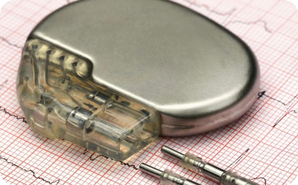

Here you find a large library of different device tracings. Browse through more than 500 tracings of different companies and different devices.