functioning of ACapTM Confirm
Tracing
Manufacturer Abbott
Device PM
N° 19
Patient
83-year-old man implanted with an Assurity MRITM pacemaker for syncopal atrioventricular block; programming of ACapTM Confirm; interrogation of the pacemaker and analysis of the automatic atrial threshold measurement curves.

Graph and trace
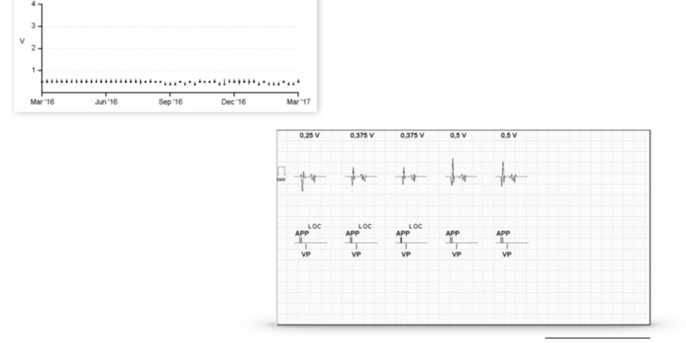
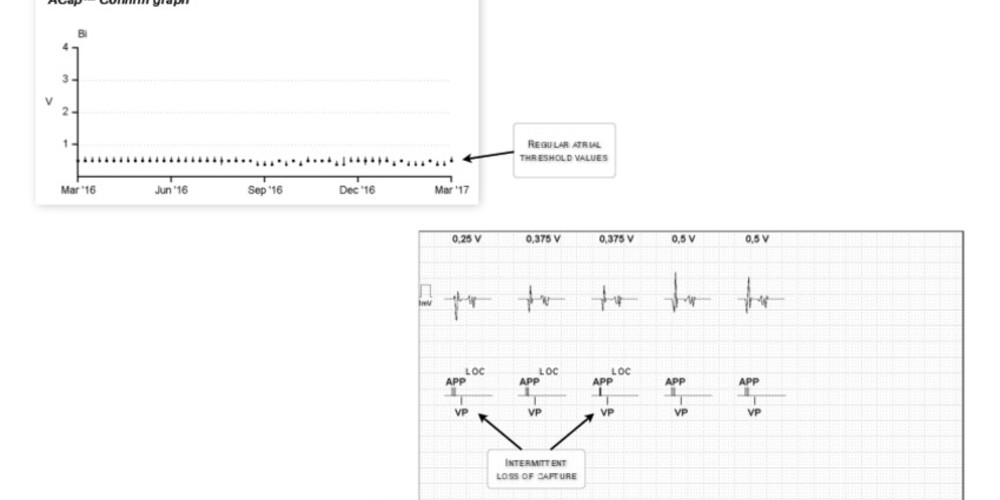
The ACapTM Confirm graph shows low (<1 Volt) and regular atrial threshold values; the tracing of the last automatic measurement shows an intermittent loss of capture at 0.25 Volts then at 0.375 Volts followed by an effective capture at 0.5 Volts which therefore constitutes the atrial threshold value.

Other articles that may be of interest to you
EGM recordings


When ACapTM Confirm is programmed to On, the atrial threshold is automatically measured every 8 or 24 hours. The measurement of the atrial threshold is based on the analysis of the morphology of the evoked atrial response. During the threshold measurement, atrial pacing pairs are delivered with a first pulse corresponding to the amplitude tested and a second 5 Volts backup pulse systematically issued 30 ms after the first capture or non-capture. To determine the morphology of the evoked response, a number of points are sampled after the first pulse and after the backup safety pulse. These points are then joined together to define the morphology of the evoked response. Three pairs of atrial pacing are delivered to test the quality of the morphology of the evoked response before each threshold measurement and to determine the morphology corresponding to a loss of capture: a first pair with pacing at 3.875 Volts (effective capture) followed by pacing at 5 Volts (non-capture because occurring during the refractory period of the atrial myocardium); a second pair with pacing at 0 Volt (non-capture) followed by pacing at 5 Volts (effective capture); a third pair with pacing at 0 Volt (non-capture) followed by pacing at 5 Volts (effective capture). The device memorizes the morphology corresponding to a failed capture. The update of the loss of capture model is performed before each threshold measurement during the Setup test. The last acquired loss of capture signal is combined with the last 15 models to compute a rolling average that is continuously updated. The algorithm uses a score to differentiate between the loss of capture morphology and the capture morphology. If the capture score is considerably different from the loss of capture score, then the setup is recommended and the pacing threshold is automatically measured.
The threshold test begins with an amplitude of 0.5 Volts above the value of the last measured threshold; every 2 cycles, the amplitude of the test pulse (systematically followed by a backup 5 Volts pulse) is decreased in steps of 0.25 Volts steps. When 2 losses of capture are observed, the amplitude of the test pulse is then increased in steps of 0.125 Volts every 2 cycles until 2 effective captures are obtained (value corresponding to the pacing threshold).
When the atrial threshold has been measured, the device adjusts the pacing amplitude for the next 8 or 24 hours depending on the programming, with a variable safety margin depending on the threshold.