Pacemaker-mediated tachycardia due to a PVC
Tracing
Manufacturer Medtronic
Device PM
Field Refractory periods, PMT
N° 22
Patient
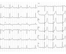
Same patient as in tracing 19; during the interrogation, recording of this episode

Graph and trace
The first line corresponds to lead I with the superimposed markers, the second line to the atrial EGM and the last line to lead II with the superimposed intervals;
- atrial sensing, ventricular pacing (AS-VP);
- PVC; far field oversensing of this PVC on the atrial channel; the AS even precedes the VS; depending on the position of the leads and the origin of the PVC, certain PVCs are detected more precociously by the atrial lead than by the ventricular lead; the spontaneous atrial activity, clearly visible on the atrial EGM, which occurs immediately after the PVC is not sensed because occurring in the post-ventricular atrial blanking;
- new PVC with oversensing;
- atrial activity, this time more remotely, is sensed (AS) and induces an AV delay and ventricular pacing; the AV delay has been automatically prolonged to meet the programmed minimum VV delay which corresponds to the programmed maximal tracking rate;
- triggering of a PMT;
- termination of the PMT by the algorithm;
- atrioventricular pacing with short AV (NCAP);
- return to sinus rhythm (AS-VP).
Other articles that may be of interest to you


PVC management is particularly difficult for a dual-chamber pacemaker. Indeed, a PVC can create a loss of atrioventricular synchrony which can, as on this tracing, induce a PMT. The PVC response algorithm is designed to prevent the tracking of retrograde P waves generated by the ventricular extrasystoles. The pacemaker defines a PVC as any sensed ventricular event that follows another ventricular event with no atrial event in the ventricular interval. The main issue with this patient is the early PVC oversensing on the atrial channel. This interval is analyzed by the pacemaker as AS-VS and is therefore not counted as a PVC. It is possible to reduce atrial sensitivity in order to prevent oversensing. Indeed, even if the atrial signal corresponding to the PVC is of considerable amplitude on the atrial EGM, it is also "softer" than a true sinus atrial signal. A change in atrial sensing may therefore correct this oversensing.